
Clinical trial registration and information
This trial is registered under NCT02658981 with preregistration date of 24 August 2016. The primary completion date was 30 April 2022. The study protocol and all related procedures were reviewed and accepted by the NCI Cancer Therapy Evaluation Program of the Division of Cancer Treatment and Diagnosis.
Ethical approval and consent
This study was approved by the Johns Hopkins Office of Human Subjects Research institutional review board (IRB), specifically IRB-2 with Committee Chair D. Cornblath. All regulatory documents were supported by the Cancer Trials Support Unit, a service of the NCI. All patients enrolled in the trial provided informed consent for tissue collection and trial enrollment as stated in the central IRB. The full list of IRB members is provided in Supplementary Information.
Patient selection
Potential participants were identified during chart review in advance of a routine clinic visit or during a routine clinic visit with a provider. Individuals were approached by the study team if willing to learn more about a study for which they may be eligible. Discussions regarding trial participation took place privately, and individuals were provided with the IRB-approved consent form. To our knowledge, there were no clear examples of self-selected bias.
Patients with histologically confirmed recurrent GBM or gliosarcoma after radiation therapy and temozolomide were recruited. Disease recurrence was defined as tumor progression despite standard-of-care treatment with the Stupp et al. protocol1. Eligibility criteria included measurable contrast-enhancing disease (≥1 cm × 1 cm on magnetic resoance imaging (MRI)) within 21 days of treatment initiation, and all patients had to be off steroids (at the equivalent dose of 7.5 mg per day of prednisone) for at least 2 weeks prior to enrollment. Last treatment of systemic chemotherapy was at least 6 weeks prior to trial initiation, and radiation or local chemotherapy with carmustine wafers was at least 12 weeks prior.
Participants had to be at least 18 years of age, have a KPS of at least 60%, have preserved organ/marrow function (including absolute lymphocyte count ≥1,000 per μl) and have no concurrent malignancies (except curatively treated basal or squamous cell carcinoma of the skin or carcinoma in situ of the cervix, breast or bladder). Prior malignancies must have been disease free on whole-body surveillance for at least 5 years. Women of childbearing potential required a negative pregnancy test and contraception use during and after treatment given risks of possible teratogenic effects with therapy. Males were also expected to use anticonception measures during treatment periods.
Exclusion criteria included any other investigational agents, active/recent autoimmune disease (except type 1 diabetes, hypothyroidism requiring only hormone replacement or non-systemic skin disorders), immunosuppressive therapy within 14 days of treatment start or >1 mg per day dexamethasone for ≥1 week before treatment. HIV-positive individuals on antiretroviral therapy were ineligible.
Study design, treatment schedule and sample collection
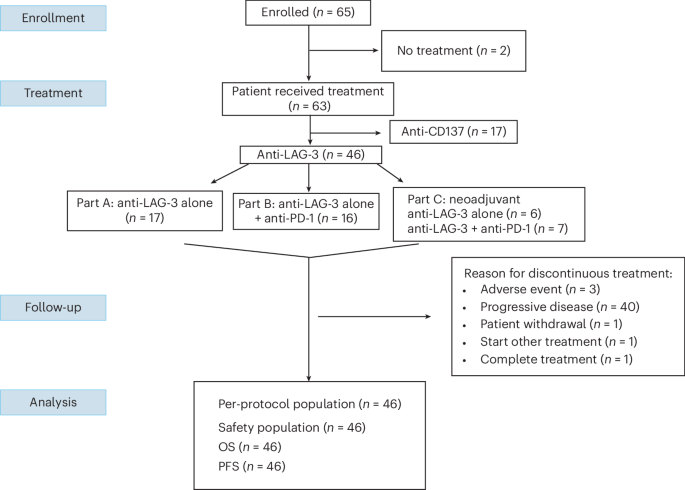
As part of the NCI ABTC, ABTC 1501 was a multicenter, phase 1 open-label platform design in the United States that allowed for multi-arm approaches with the opportunity to add on or drop off checkpoint agent/agents during the trial (Table 1). The initial study agents were anti-LAG-3 and anti-CD137 mAb (BMS-663513) administered as a single agent or in combination with nivolumab antibody for patients with recurrent GBM. Any patients deemed surgical candidates underwent maximally safe surgical resection prior to initiation of therapy. At time of surgery, patient samples were collected and stored in either saline or formalin for pathology processing per standard of care and downstream immunophenotyping. For the purposes of this paper, we focused on the relatlimab portion of the study and will report findings involving anti-CD137 antibodies in a subsequent report. The study was designed in three parts for safety evaluation with a target DLT rate of less than 30%.
Part A determined the MTD of relatlimab monotherapy in patients who were assigned either relatlimab or anti-CD137 monotherapy by sequential allocation as designed by our statistical team. A modified rule-based dose escalation with a total of seven patients was required per dosage cohort to determine the MTD. There were three prespecified doses of relatlimab at 80 mg, 240 mg and 800 mg. These dosages were based on studies in other cancers treated with the same investigational agents30.
Part B aimed to identify a safe combination regimen of relatlimab when administered in combination with nivolumab. Treatment with the combination of relatlimab and nivolumab was initiated after the MTD of relatlimab was confirmed in part A. Nivolumab dosages followed historical safe dosages (240 mg). Given increased rates of immune-related adverse events with combination immunotherapy31, the plan was to originally dose relatlimab one step down from the MTD that was determined in part A. However, due to reports of increased rates of myocarditis with combination nivolumab and relatlimab in another trial13, the decision was made to start relatlimab dosage at 80 mg and escalate to 160 mg for combination therapy with 240 mg nivolumab. Patients again were assigned by sequential allocation.
Part C examined the safety and impact of neoadjuvant relatlimab monotherapy and combination relatlimab/nivolumab therapy on the immune microenvironment for recurrent GBM. Patients received one preoperative (neoadjuvant) dose 10 days (±3 days) prior to planned surgery. After surgery, patients underwent treatment with either relatlimab alone (800 mg) or combination relatlimab (160 mg every 2 weeks) and nivolumab (240 mg every 2 weeks). Sample sizes for parts A, B and C were determined based off of previous trials for GBM (Extended Data Table 1) and published FDA guidance for expansion cohorts32. All procedures were approved by the Cancer Therapy Evaluation Program with a central IRB and Brain Malignancy Steering Committee as above, with all patients enrolled in the clinical trial consented for tissue specimen and blood collection for research. Researchers involved with tissue processing and immune assays were blinded to patient demographics.
Clinical assessments
The safety evaluation period for dose-escalation decisions was 4 weeks from the initial dose (cycle 1). Version 5.0 of the NCI CTCAE was used for scoring toxicity and adverse events. Safety assessments involved monitoring and recording all adverse events, pregnancy testing (in women with childbearing potential), serial monitoring of hematology and blood chemistry, regular measurement of vital signs and physical/neurological examinations; electrocardiograms and other cardiac monitoring were performed as necessary after clinical evaluation. All adverse events were evaluated and recorded during the trial period. Patients with measurable disease were assessed by MRI prior to every odd cycle.
mRANO criteria
Patients with measurable (1 cm × 1 cm) contrast-enhancing disease at baseline were assessed by MRI prior to every odd cycle of immune checkpoint inhibitor treatment under mRANO criteria14 to mitigate potential immunotherapy-related pseudoprogression by requiring confirmation of radiographic progression within a 6-month window of starting treatment in non-surgical patients. Complete response was defined as disappearance of all enhancing and non-enhancing disease for at least 4 weeks, no new lesions, discontinuation of corticosteroids (except physiologic doses), stable or improved T2/FLAIR lesions and stable or improved clinical status.
Partial response required at least 50% reduction in the sum of perpendicular diameters of measurable lesions sustained for 4 weeks, no progression of non-measurable disease, no new lesions, corticosteroid dose no greater than baseline and stable or improved T2/FLAIR lesions and clinical status; again, non-measurable disease precludes partial response. Stable disease represents maintenance of status that does not qualify for complete response, partial response or progression, requiring 4-week duration with stable imaging findings on same or lower corticosteroid doses.
Progressive disease was defined by at least 25% increase in tumor measurements, significant increase in T2/FLAIR lesions not due to other causes, any new lesion, clear clinical deterioration not attributable to other factors, death or clear progression of non-measurable disease. For the first 6 months after initiation of treatment, radiographic progression required confirmation of progression as defined by increased contrast-enhancing tumor growth more than 2 months after initial progressive enhancement to be considered progressive disease. Pseudoprogression was defined as initial progressive enhancement within the first 6 months of treatment followed by disease stability or regression on the confirmation scan. If radiographic progression occurred after 6 months from the start of treatment, no confirmation was required for progressive disease.
Immune correlation studies
Immunofluorescence
An automated multiplex immunofluorescence (mIF) panel was developed for use in FFPE GBM human samples using methods previously described33. The final 3-plex panel was validated on archival GBM to show equivalence with serial sections of individual immunohistochemistry (IHC) stained slides.
In brief, samples were baked offline for 3 h at 65 °C and loaded onto a Leica BOND RX automated research stainer (Leica Biosystems). Samples were baked online at 60 °C for 30 min, and residual paraffin was removed (Dewax; Leica Biosystems). Initial antigen retrieval was performed using a pH 9 EDTA buffer (ER2; Leica Biosystems) for 40 min at 100 °C, followed by another round of antigen retrieval using pH 6 sodium citrate buffer (ER1; Leica Biosystems) at 95 °C for 20 min. After initial blocking for endogenous peroxidases (BLOXALL; Vector Labs), non-specific antibody binding was blocked (Protein Block; Agilent Technologies). All primary antibodies were diluted in antibody diluent with background-reducing components (Agilent Technologies). Primary antibodies, polymers and opals were applied for Position 1 (see table), and then antibody stripping was performed using a pH 6 sodium citrate buffer (ER1; Leica Biosystems) for 20 min at 95 °C. This process was repeated for each position, after which slides were counterstained (Spectral DAPI; Akoya Biosciences) and coverslipped (ProLong Diamond; Invitrogen).
Whole-slide scans of the mIF-stained slides were acquired using PhenoImager HT (Akoya Biosciences) with the standard set of multispectral slide scan filters. Regions of interest were selected using Phenochart version 1.1.0 (Akoya Biosciences) and then unmixed using a microscope-specific library consisting of the matching fluorophores using inForm version 2.5.1 (Akoya Biosciences). Individual unmixed TIFF files were stitched together using HALO version 6.8 (Indica Labs). Using this software, we quantified the percentages of cells positive for CD8, PD-1 and/or LAG-3. Due to substantial intratumoral heterogeneity of GBM, CD8+ T cells, CD8+PD-1+, CD8+LAG-3+ and CD8+PD-1+LAG-3+, subsets were quantified across entire whole-slide images rather than within predefined regions of interest. All measurements were obtained from a single experimental replicate (n = 1) and a single experimental run to minimize batch effects and ensure analytical consistency. Each dot represents whole-slide data from a single patient.
RNA isolation and gene expression profiling
Total RNA was extracted from 5-µm-thick FFPE sections obtained from surgical resection of patients with recurrent GBM using an RNeasy FFPE Kit (Qiagen) following the manufacturerʼs protocol. RNA was quantified using Qubit and NanoDrop. Then, 100 ng of total RNA was run on an nCounter SPRINT Profiler system for gene expression profiling using the commercial human pan-cancer immune profiling panel with 770 immune-related genes, including 14 internal reference controls (XT-PGX-Huv1, NanoString; Bruker Spatial Biology). Data were analyzed using nSolver 4.0 software with an nCounter Advanced Analysis 2.0 add-on (NanoString; Bruker Spatial Biology).
T cell clonality
Genomic DNA from FFPE samples were extracted (Qiagen AllPrep DNA/RNA Kit) and amplified by polymerase chain reaction (PCR) with primers (Adaptive Biotechnologies) that span Vβ and Jβ regions to capture TCRβ gene segments. Sequencing adapters were ligated using a library preparation kit and performed on an Illumina platform (MiSeq), and raw sequencing reads were processed (MiXCR) to identify clonotypes and their relative frequencies. Simpson clonality was calculated to assess the TCR repertoire with visualization and statistical analysis performed using GraphPad Prism.
Gene expression profiling
FFPE samples from surgical resection of patients with recurrent GBM were deparaffinized for RNA extraction. Reporter and capture probes for genes of interest were mixed in hybridization buffer with the sample at 65 °C for 16–20 h with hybridized samples loaded into nCounter cartridge and nCounter Digital Analyzer for barcode detection to measure gene expression (NanoString; Bruker Spatial Biology). Results were analyzed using nSolver (NanoString; Bruker Spatial Biology). P values for differential expression were computed using a negative binomial generalized linear model with Wald test statistics in the NanoString nSolver Advanced Analysis module. In this exploratory analysis, significance was defined as a nominal P < 0.05.
MIBI-TOF
MIBI-TOF was performed using a custom human panel targeting immune (CD45, CD4, CD8, Foxp3, P2RY12, CD68, CD14, CD20, HLA1, CD163, CD206), tumor (Sox9, Olig2), exhaustion (PD-1) and metabolic (ASCT2, CD36) markers, obtained in a preformulated, lyophilized format from IONpath, Inc. Antibodies were resuspended in TBS-IHC Tween with 3% donkey serum and filtered before staining. Tissue preparation followed standard protocols, including deparaffinization, antigen retrieval, blocking, overnight antibody incubation, fixation and dehydration. Hematoxylin and eosin (H&E)-stained sections were reviewed for quality control, and background staining was manually assessed.
MIBI data were acquired using a uniform field of view (800 µm2, when possible), an 8.5-nA beam current and a 0.58-ms dwell time. Background correction and noise removal settings were turned off. To correct for signal contamination, we applied Rosetta Matrix Compensation, a method that subtracts known overlapping markers using an empirically determined matrix34. Intensity normalization was performed using median pulse height (MPH), fitting a polynomial function to mass-specific data across runs and adjusting signal drift based on a predefined normalization curve34.
Cells were segmented using the deep-learning-based Mesmer model, using HH3/dsDNA for nuclear segmentation and a composite of CD14, CD45, CD8 and HLA1 for membrane segmentation34. Cell phenotyping was conducted using Pixie, an unsupervised pixel clustering algorithm that overclustered pixels and cells before hierarchical consensus clustering into 20 metaclusters35,36,37. Assignments were validated through manual inspection in Adobe Photoshop, and systematic errors were corrected by refining clustering parameters.
Statistical analysis
Baseline patient and disease characteristics were summarized using descriptive statistics. Safety data were evaluated based on NCI CTCAE version 5, and the incidence rate of adverse events was presented using a proportion of the total number of treated patients for each group. Tumor responses were initially classified by investigator assessment and subsequently followed by a central review. PFS and OS were measured from date of treatment initiation to database locking, with exceptions for clinical event occurrence or censorship. Survival probability was estimated using the Kaplan–Meier method. The confidence interval of median survival time was constructed by the Brookmeyer–Crowley method. Immune correlative studies were exploratory and mechanistic in nature, and the data were presented either descriptively or through two-tailed Studentʼs t-test, with P < 0.05 considered statistically significant using R and GraphPad Prism. The analysis of clinical data was performed using SAS version 9.4 (SAS Institute).
Reporting summary
Further information on research design is available in the Nature Portfolio Reporting Summary linked to this article.



