
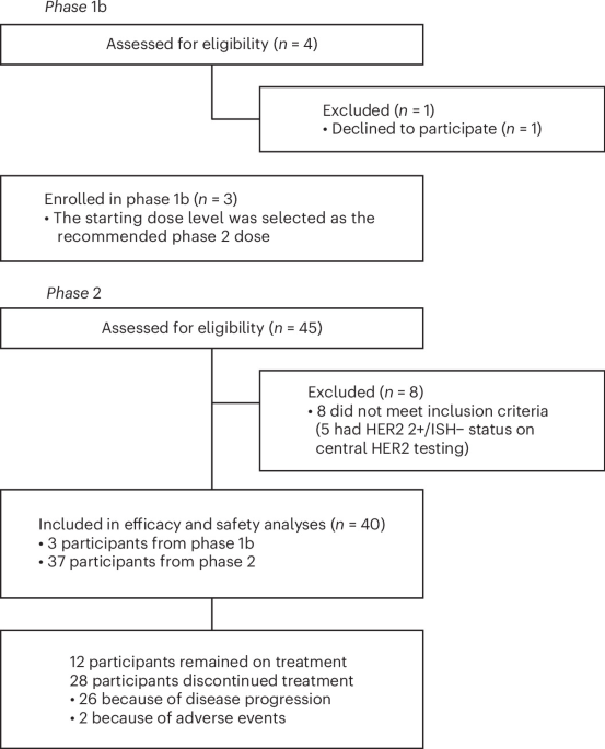
Trial design and participants
The HERBOT study was an open-label, single-arm, phase 1b/2 trial performed at 12 academic cancer centers in South Korea to evaluate the efficacy and safety of a quadruplet regimen (trastuzumab, nivolumab, gemcitabine and cisplatin) as first-line therapy for HER2-positive unresectable advanced BTC. Adults aged ≥20 years with HER2-positive histologically confirmed unresectable, locally advanced or metastatic BTC including intrahepatic or extrahepatic cholangiocarcinoma and gallbladder carcinoma, were eligible for inclusion. Eligible participants included those with previously untreated disease that was unresectable or metastatic at initial diagnosis and those who developed recurrent disease >6 months after surgery with curative intent and >6 months after the completion of adjuvant therapy. Up to one cycle of gemcitabine-based chemotherapy for advanced, unresectable or metastatic cholangiocarcinoma before enrollment was allowed. Other inclusion criteria included an ECOG performance status of 0 or 1, measurable lesion per RECIST version 1.1 and adequate organ function. Participants were excluded if they had multiple primary cancers, active or prior documented autoimmune or inflammatory disorders, previously received ICIs or known allergy or hypersensitivity to any of the study treatments. Full inclusion and exclusion criteria can be found in the online protocol. Participants were enrolled regardless of sex, which was obtained from the medical record at enrollment.
Ethical approval and consent
The trial was conducted in accordance with the Declaration of Helsinki and the International Council for Harmonization Guidelines for Good Clinical Practice (ClinicalTrials.gov identifier: NCT05749900). The trial protocol was approved by the Korean Cancer Study Group Protocol Review Committee, the institutional review board of Severance Hospital, Yonsei University Health System and the institutional review board at each participating institution (Extended Data Table 5). All participants provided written informed consent before enrollment.
Procedures
Participants were enrolled on the basis of centrally confirmed HER2 positivity, defined as IHC 3+ or IHC 2+ combined with ISH+ or ERBB2 gene copy number ≥ 6.0 by next-generation sequencing tested in a Clinical Laboratory Improvement Amendments-certified laboratory at Severance Hospital. Before the study, tumor tissues were obtained from either primary or metastatic tumors (archival or fresh). To determine HER2 status, an anti-HER2/neu antibody (clone 4B5; Ventana Medical Systems) was used for IHC; the HER2 expression scoring system was applied using the guideline26. SISH was performed using INFORM HER2 DNA and chromosome 17 (CEP17) probes (Ventana Medical Systems). HER2 amplification was defined as a HER2/CEP17 ratio ≥ 2.0 or an average HER2 gene copy number ≥ 6 signals per cell.
Phase 1b had a 3 + 3 design with dose level 0 and level −1. The starting dose (dose level 0) was intravenous nivolumab 360 mg on day 1, intravenous trastuzumab (Samfenet, Samsung Bioepis) 6 mg kg−1 (after 8 mg kg−1 load) on day 1, intravenous gemcitabine 1,000 mg m−2 on days 1 and 8 and intravenous cisplatin 25 mg m−2 on days 1 and 8 every 3 weeks; dose level −1 was a reduced dose of gemcitabine (800 mg m−2). Dose-limiting toxicities were assessed during the first treatment cycle of the phase 1b portion according to the protocol-defined hematologic and nonhematologic toxicity criteria. In phase 2, the RP2D from phase 1b was used. All participants were treated until documented disease progression, death, unacceptable toxicity or up to nine cycles for gemcitabine and cisplatin and up to 24 months for nivolumab and trastuzumab (chemotherapies beyond ninth cycles were permitted per the investigator’s discretion). Computed tomography or magnetic resonance imaging was performed within 28 days before the treatment’s first dose as the baseline; tumor responses were evaluated every three cycles according to the RECIST version 1.1 criteria. Safety and tolerability were assessed from the study treatment’s first dose at each clinic visit until the treatment ended. All treatment-emergent adverse events were collected and graded on the basis of the National Cancer Institute Common Terminology Criteria for Adverse Events 5.0. Investigators assessed the relationship of each event to study treatment. Left-ventricular ejection fraction was measured using a multigated acquisition scan or echocardiography at baseline and repeated every 12 weeks. Following treatment discontinuation, participants were monitored for survival every 3 months until death, loss to follow-up, consent withdrawal or study termination. Quality of life was assessed before treatment and on day 1 of every six cycles till the end of treatment, using the European Organization for Research and Treatment of Cancer Quality of Life Questionnaire C30 participant-reported outcomes, which will be reported separately. Protocol-based blood samplings were prospectively performed at baseline, at cycle 2 day 1, at cycle 4 day 1 and at progression.
Clinicopathologic feature analyses
We collected data on the following variables: age at the beginning of treatment, sex, ECOG performance status, pathology, plasma levels of albumin, lactate dehydrogenase, CEA and CA 19-9, neutrophil and lymphocyte counts and metastatic organs. Parameters that were previously reported to be related to prognosis—ECOG performance status, neutrophil-to-lymphocyte ratio and tumor markers—were additionally analyzed34.
Outcomes
This trial’s primary endpoints were determination of the RP2D for phase 1b and ORR according to RECIST version 1.1 for phase 2. Secondary endpoints included PFS, OS, DOR, DCR, safety profile and exploratory biomarker analyses. The ORR was calculated as the percentage of participants who achieved a confirmed CR or PR, with confirmatory tumor assessment required at the next scheduled evaluation or at least 4 weeks after the initial response assessment. DCR was the proportion of subjects with confirmed CR, PR and stable disease for at least 4 weeks. PFS was the time from treatment initiation to the date of disease progression or death from any cause. For participants with no disease progression or who did not die, the censoring date was the last date at the nearest time of their last response evaluation. OS was the time from treatment initiation to the date of death from any cause. The OS censoring date was the last date the subject was known to be alive. In cases where radiologic progressive disease (PD) was considered by the investigator to represent possible pseudoprogression related to immunotherapy, treatment could be continued until PD was confirmed by repeat tumor assessment performed within 4–8 weeks, in accordance with iRECIST criteria35.
Preplanned exploratory analyses
The analysis of HER2-stained WSIs was performed using Lunit SCOPE HER2, which is an HER2 IHC-specific AI model trained to perform cancer tissue segmentation and detection of tumor cells. The model was trained on 2,966 WSIs from multiple cancer types including BTC and gastric cancer, encompassing a total tissue area of 29,589.12 mm2 and 2,509,177 cells annotated by board-certified pathologists. The model performed cell classification (HER2 0, 1+, 2+ and 3+) according to IHC membrane staining intensity. On the basis of the model-detected results, a slide-level proportion of HER2 3+ tumor cells was calculated.
H&E-stained WSIs were analyzed using Lunit SCOPE IO, a previously validated AI-based analyzer, to classify tumor immune phenotypes. The model was developed across 26 different tumor types, using annotations from board-certified pathologists of 2,828,448 cells and 1.07 × 1010 µm2 of cancer areas and stromal regions. For each H&E WSI, Lunit SCOPE IO performs tissue segmentation into cancer or stroma areas and executes cell detection to identify tumor cells and lymphocytes. The WSI is then partitioned into 0.25-mm2 grids, where grid-level immune phenotypes are determined on the basis of specific lymphocyte density thresholds. The IIP is defined by an intratumoral lymphocyte density ≥ 130 per mm2, while the IEP is defined by an intratumoral lymphocyte density < 130 per mm2 and stromal lymphocyte density ≥ 260 per mm2. Grids with lymphocyte densities falling below these thresholds in both regions are classified as IDP. The final representative slide-level immune phenotype is determined by the proportion of these grids. A slide is categorized as inflamed if the proportion of inflamed grids is ≥33.3%. If the inflamed criteria are not met, the slide is classified as IEP if the proportion of immune-excluded grids is ≥33.3% or IDP if it fails to meet either threshold.
Statistical analyses
The sample size was calculated on the basis of the primary outcome of ORR using Simon’s two-stage minimax design. Previous data from the TOPAZ-1 study informed the null hypothesis and our assumption was that the regimen would be considered inactive if the ORR was ≤27%, whereas ≥45% was considered indicative of sufficient activity (P0 = 0.27, P1 = 0.45). On the basis of a one-sided, type I error rate of 10% and a power of 80%, 34 evaluable participants were required for the phase 2 portion of the study. The first stage required 16 participants and the study was to be terminated for futility if fewer than five objective responses were observed. The regimen would be considered worthy of further investigation if at least 14 objective responses (corresponding to an ORR of 41.2%) were observed among the 34 evaluable participants. To account for an anticipated dropout rate of approximately 10%, the planned phase 2 accrual target was 38 participants. The protocol planned to enroll approximately 41–44 participants across the phase 1b and phase 2 portions of the study.
The primary efficacy analysis was performed in the full analysis set (FAS), defined as all participants who received at least one dose of study treatment and had at least one baseline tumor assessment. Safety analyses were performed in all participants who received at least one dose of study treatment. All participants who received the RP2D, including those enrolled in phase 1b, were to be included in the FAS for efficacy and safety analyses. ORR and DCR were reported as proportion and with two-sided 95% exact CIs using the Clopper–Pearson method based on binomial distribution. The Kaplan–Meier method, assuming noninformative censoring, was used to estimate median time-to-event endpoints (PFS, OS and DOR) with associated 95% CIs using the Brookmeyer and Crowley method. Kaplan–Meier estimates of the survival function along with 95% CIs at specific time points were summarized and their standard errors calculated using Greenwood’s formula. In post hoc subgroup analyses, we investigated the association between baseline participant characteristics and the median PFS and OS. Survival differences were assessed using the log-rank test and HRs were estimated using Cox proportional hazards models based on Wald tests, with ties handled using the Breslow method. The proportional hazards assumption was verified with Schoenfeld residuals. A post hoc landmark analysis was performed using 6.2 months (corresponding to the median time to complete nine cycles of treatment) as the landmark time, with both the PFS and OS landmark analyses including only participants who remained alive and progression-free at the landmark time. No adjustments for multiple testing were applied in the post hoc subgroup analyses because of their exploratory and hypothesis-generating nature.
Participant baseline characteristics are summarized using descriptive statistics, with categorical variables expressed as frequencies and percentages and continuous variables expressed as medians (IQR). Comparisons between groups for categorical variables were performed using the chi-square test or Fisher’s exact test, as appropriate. Linear regression was used for visualization of the relationship between two continuous variables, with the fitted regression line presented together with the 95% CIs (shown as a shaded gray band). Correlation coefficients were calculated using Pearson’s or Spearman’s method, depending on data distribution, with normality assessed using the Shapiro–Wilk test.
All P values were two-sided, with P < 0.05 indicating statistical significance. No sensitivity or interim analysis was planned. All statistical analyses and visualizations were performed using R software (version 4.4.0; R Foundation for Statistical Computing) and SAS software (version 9.4; SAS Institute).
Reporting summary
Further information on research design is available in the Nature Portfolio Reporting Summary linked to this article.



