
Trial design and oversight
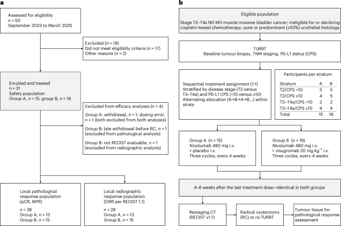
GDFATHER-NEO (NCT06059547; EudraCT 2022-002816-22; EU CT 2024-513957-55-00) is an exploratory, stratified, participant-blinded, parallel-group phase 2a trial of neoadjuvant PD-1 blockade with or without GDF-15 neutralization in patients with MIBC who are ineligible for or who decline cisplatin-based chemotherapy. Before allocation, patients were stratified by clinical tumor stage (T2 versus T3–T4a) and by PD-L1 expression on tumor and immune cells, determined by local pathology as a combined positive score (CPS ≥10 versus <10), into one of four strata: T2 and CPS <10, T2 and CPS ≥10, T3–T4a and CPS <10, and T3–T4a and CPS ≥10 (Fig. 1b).
Within each stratum, patients were allocated 1:1 to nivolumab plus visugromab (group B) or to nivolumab plus placebo (group A). Allocation was performed centrally and sequentially within each stratum (A–B–A–B, …) until at least 12 efficacy-evaluable patients had been allocated to each group. Sequential allocation with blinding of patients, rather than randomization, was chosen because of the small size of the individual strata, to prevent imbalance between treatment arms, and because this was the first evaluation of combined GDF-15 and PD-1 blockade in patients with early-stage disease treated with curative intent. Investigators and sponsor staff were aware of treatment allocation; patients were not. Local pathologists and the independent central pathology and imaging reviewers assessed responses without knowledge of treatment allocation.
The trial was sponsored by CatalYm GmbH (Munich, Germany). The protocol and its amendments were approved by the competent ethics committee of the coordinating center (Comitato Etico Territoriale Lombardia 1) and by the Italian regulatory authority (Agencia Italiana del Farmaco), and the trial was conducted in accordance with the principles of the Declaration of Helsinki, the guidelines for Good Clinical Practice of the International Council for Harmonisation and applicable local law. All patients provided written informed consent before any trial-specific procedure and received no compensation for participation.
The clinical cutoff date was 15 July 2026 for the analyses reported here and was supplemented by central review, bioanalytical and translational data. Data management, biometrics, on-site monitoring and bioanalytical and translational analyses were performed by, or under the oversight of, the sponsor. Independent central review of surgical specimens (pathologic response) and of imaging (radiographic response) was performed by Discovery Life Sciences Biomarker Services GmbH and by Median Technologies, respectively.
Participants
Patients aged ≥18 years with newly diagnosed cT2–T4a N0 M0 MIBC who were ineligible for cisplatin-based chemotherapy according to the modified Galsky criteria29 or who declined such therapy, and who were considered fit to undergo radical cystectomy after 12 weeks of neoadjuvant therapy, were eligible. Clinical staging by CT was mandatory; positron emission tomography (PET)–CT and multiparametric MRI (mpMRI) were optional. Archival tumor material from a TURBT performed within 3 months before the start of treatment had to be available as a paraffin block. Only patients with an Eastern Cooperative Oncology Group performance status of 0 or 1 were eligible.
Further inclusion criteria were adequate bone marrow function (haemoglobin ≥10 g dl−1, platelet count ≥100 × 109 l−1, leukocyte count ≥2.5 × 10⁹ l−1), adequate hepatic function (aspartate and alanine aminotransferase ≤2× the upper limit of normal (ULN); bilirubin ≤1.5 × ULN), adequate renal function (serum creatinine <1.5× ULN and/or creatinine clearance ≥50 ml min−1) and coagulation parameters within the normal range. Patients with type 2 diabetes mellitus had to discontinue metformin, which alters circulating GDF-15 concentrations, and switch to an alternative antidiabetic agent before the start of treatment.
Patients who had received prior systemic anticancer therapy, prior radiotherapy to the bladder tumor or a partial cystectomy, and patients who declined radical cystectomy at the time of trial entry were excluded. Patients with major tissue injury, clinically significant cardiovascular disease, active autoimmune disease, chronic high-dose systemic corticosteroid treatment or known or expected hypersensitivity to visugromab or nivolumab were also excluded. Full eligibility criteria are provided in the trial protocol (Supplementary Information). Sex was recorded as documented in the medical record and was used for descriptive purposes only; because of the small sample size, no sex- or gender-based analyses were prespecified, and none were performed.
Surgical procedures
A TURBT performed according to local practice within 3 months before the start of treatment was required for every patient, for histological characterization and staging. The resulting paraffin-embedded tumor material had to be made available for the additional histological and molecular analyses of the trial, in particular for PD-L1 CPS scoring; residual material was returned to the providing pathology department on request.
The trial comprised a screening phase of up to 4 weeks, beginning on the day written informed consent was obtained, and a core phase of 19 weeks, beginning on the day of the first administration of trial treatment. The core phase comprised the three 4-week immunotherapy cycles and a window of 4–8 weeks after the last neoadjuvant dose during which surgery was to be performed. Radical cystectomy or re-TURBT was planned at the time of trial entry and provided the surgical specimen (paraffin block) required for the assessment of pathologic response.
After completion of neoadjuvant treatment, and at the discretion of the investigator and the patient, one of the following procedures was performed, in line with the recommendations of the European Association of Urology: RC with pelvic lymph node dissection (PLND); or maximal re-TURBT, with adjuvant chemoradiotherapy or adjuvant nivolumab to be considered as clinically appropriate.
Provision of tumor material for the assessment of pathologic response and of immune cell infiltration was mandatory irrespective of whether a radical or a bladder-sparing approach was chosen. Surgical procedures followed each center’s standard practice. Patients who declined, or who were unsuitable for, RC or re-TURBT after neoadjuvant treatment were managed according to European Association of Urology guidelines, which recommend chemoradiotherapy as the principal organ-sparing option. An end-of-treatment (EoT) visit was scheduled 21 days (±2 days) after completion of the surgical procedure.
Neoadjuvant treatment and procedures
Visugromab (CTL-002) is an investigational, humanized, hinge-stabilized immunoglobulin G4 (IgG4) monoclonal antibody that binds GDF-15 with high affinity. It was administered intravenously over approximately 60 min every 4 weeks (Q4W) at 20 mg kg−1. Pharmacokinetic–pharmacodynamic modeling of data from the first-in-human trial indicated that, in combination with nivolumab, complete neutralization of circulating GDF-15 is achieved with 10 mg kg−1 every 2 weeks or with 20 mg kg−1 every 3–4 weeks.
Nivolumab, a fully human IgG4 monoclonal antibody directed against PD-1, was administered in accordance with its summary of product characteristics30 and local guidelines. The 480 mg Q4W schedule was selected so that both agents could be given on the same 4-weekly visit schedule (Fig. 1b).
In group B, nivolumab and visugromab were given on day 1 of each of the three cycles, with visugromab administered first, and followed by a 30-min observation period before the nivolumab infusion. In group A, patients received placebo (drug-free infusion bags identical in appearance to the visugromab bags) before nivolumab, on the same schedule.
AEs were graded according to the National Cancer Institute Common Terminology Criteria for Adverse Events version 5.031 and were managed according to toxicity-specific algorithms defined in the protocol. Guidance on optional premedication before visugromab and on dose interruption of nivolumab and visugromab in the event of immune-related AEs was provided to investigators in the protocol (Supplementary Information).
Clinical assessments and outcomes
The primary objective was to assess the antitumor activity of visugromab in combination with nivolumab. Secondary objectives were to assess the safety and tolerability of the combination, its pharmacokinetics and pharmacodynamics, the development of antidrug antibodies (ADAs), and clinical outcomes in the neoadjuvant setting. Collection of molecular and genomic profiling data from peripheral blood and tumor tissue was an exploratory objective. Optional 18F-fluorodeoxyglucose PET–CT and mpMRI were permitted for exploratory response assessment.
The two primary endpoints were the rate of pCR, determined by blinded local pathologists, and the rate of radiographic response according to RECIST version 1.132, determined by investigators. Secondary endpoints were AEs and serious AEs (SAEs), ADAs, delay of surgery, the rate of major pathologic response (MPR), event-free survival (EFS), time to relapse and overall survival (OS), standard pharmacokinetic parameters, baseline serum GDF-15 concentrations, and pCR, MPR and radiographic response as determined by independent central review.
pCR was defined as the absence of any residual viable tumor in the RC or re-TURBT specimen. Where a re-TURBT was followed by an RC, the pathologic evaluation of the RC specimen was used. Residual non-muscle-invasive disease (ypTa or ypT1) and residual carcinoma in situ were not considered a pCR. MPR was defined as ypT0/Ta/Tis/T1 N0 M0 after RC or ypT0/Ta/Tis/T1 NX M0 after re-TURBT, and therefore includes pCR. Radiographic response rate was defined as the proportion of patients with a complete or partial response according to RECIST version 1.1. Four protocol amendments took place. One implemented clarifications requested by local authorities (Italy) before the start of patient enrolment (removal of need for mandatory hospitalization after first administration of study medication in cycle 1 and extension of screening period to 28 days). A second amendment introduced re-TURBT as a surgical alternative to RC together with the assessment of ADAs as an additional secondary endpoint. A third amendment allowed inclusion of participants with stable atrial fibrillation. Post-enrollment, by the fourth amendment, central pathological review (for pCR) and central radiographic review (for ORR) were formally established as secondary endpoints (complementary central review had been mentioned in early protocol versions already) in line with clarifications on definitions of pCR and MPR.
A safety follow-up visit was scheduled 90 days (±3 days) after the last neoadjuvant dose to capture all AEs and SAEs occurring after neoadjuvant treatment, including postoperative complications. Longer-term outcomes were captured at follow-up visits for up to 12 months after RC or re-TURBT.
Exploratory biomarker endpoints, serum visugromab and ADA measurements
For the assessment of total visugromab, serum samples were collected 30 min before infusion and immediately before the end of infusion during cycles 1–3, on days 2 and 8 of cycle 1 and at the EoT visit. For the assessment of ADAs, serum samples were collected 30 min before infusion on day 1 of cycles 1–3 and at the EoT visit. Serum was isolated using standard procedures, cryopreserved at −80°C and analyzed using validated electrochemiluminescence assays for total visugromab and for ADA detection.
Formalin-fixed, paraffin-embedded (FFPE) tumor tissue obtained at screening (TURBT) and at post-treatment RC or re-TURBT was sectioned at 4 μm. After histological confirmation of tumor content on H&E-stained sections, multiplex immunofluorescence was performed using a validated 6 + 1-plex OPAL assay (Akoya Biosciences) on a Leica Bond Rx automated staining platform (BOND-RX software version 7.0.3.216, Leica Biosystems). The panel comprised CD4 (F7.2.38, Cell Marque), CD8 (C8/144B, Agilent), Ki-67 (MIB-1, Agilent), FoxP3 (SP97, Abcam), PD-L1 (SP263, Roche) and GDF-15 (HPA011191, Sigma-Aldrich). Chromogenic immunohistochemistry for pan-cytokeratin (AE1/AE3, Agilent) was subsequently performed on the same slide using a Ventana Discovery Ultra platform (software version 12.5 or higher) to enable segmentation of tumor compartments.
Whole-slide multispectral fluorescence imaging was performed at 20× magnification using an Akoya PhenoImager HT/Vectra Polaris platform (software version 1.0.13), followed by bright-field scanning after pan-cytokeratin staining. Pathologist-assisted digital image analysis was performed using validated Visiopharm algorithms (version 2021.09.2.11085, Visiopharm). Tumor, stromal and adjacent non-neoplastic tissue compartments were segmented on the basis of pan-cytokeratin staining, and predefined immune cell and biomarker-positive populations were quantified as cell densities (cells mm−2) and as relative proportions within each compartment. Staining quality was assessed using on-slide and internal tissue controls, and markers that failed predefined quality criteria were excluded from downstream analyses.
Visiopharm assigns each detected cell to a single, mutually exclusive phenotype; cells co-expressing several markers (for example, CD8+Ki-67+) are therefore not included in the corresponding single-positive populations. To derive total marker-positive cell densities, composite variables were reconstructed by summing all mutually exclusive phenotypes containing the marker of interest. All reconstructed densities were calculated within the same tissue compartment and are expressed as cells per square millimeter.
All assays were performed according to predefined analytical procedures and validation criteria established for exploratory profiling of the tumor microenvironment and of GDF-15 biology.
Evaluation of intratumoral pro-GDF-15 expression in human FFPE samples
Intratumoral pro-GDF-15 expression was determined using a rabbit polyclonal anti-GDF-15 antibody (HPA011191, Sigma-Aldrich) as the primary antibody, with automated staining on a Ventana BenchMark Ultra platform (Roche; software versions 12.3.1 and 12.5.4). Binding of the anti-GDF-15 antibody was visualized using a secondary horseradish peroxidase-conjugated antibody (Ventana OptiView Universal DAB Detection Kit, 760-700; Roche Diagnostics) and a precipitating enzyme reaction product. Slides were evaluated by a board-certified pathologist who scored cytoplasmic staining of tumor cells and, where applicable, membranous staining. Cytoplasmic staining was assigned to four intensity categories, from 0 (no staining) to 3+ (intense staining), and the percentage of stained cells in each category was recorded. The H score, which reflects tumor GDF-15 protein levels, was calculated by summing the products of the percentage of cells in each category and the corresponding intensity value, as follows:
$$\begin{array}{l}{\rm{H}}\; {\rm{score}}=[1\times ({\rm{percentage}}\; {\rm{of}}\; {\rm{intensity}}1)]\\\qquad\qquad+[2\times ({\rm{percentage}}\; {\rm{of}}\; {\rm{intensity}}2)]\\\qquad\qquad+[3\times ({\rm{percentage}}\; {\rm{of}}\; {\rm{intensity}}3)]\end{array}$$
Central evaluation of PD-L1 protein expression in human FFPE samples
PD-L1 expression was assessed centrally by immunohistochemistry using the SP263 assay (790-4905; Roche Diagnostics) according to the manufacturer’s protocol, with a rabbit IgG monoclonal antibody (790-4795; Roche Diagnostics) as isotype control. Staining was evaluated by a board-certified pathologist, and the tumor proportion score (TPS) and CPS were determined as previously described.
Measurement of cytokine and chemokine levels in patient samples
Serum samples for cytokine and chemokine analysis were collected 30 min before infusion on day 1 of cycles 1–3 and at the EoT visit. CXCL9 (MIG) and CXCL10 (IP-10) concentrations were measured using validated ELISAs (Human CXCL9/MIG Quantikine ELISA Kit, DCX900; Human CXCL10/IP-10 Quantikine ELISA Kit, DIP100; R&D Systems). IFN-γ concentrations were determined using a validated multiplex immunoassay (V-PLEX Plus Proinflammatory Panel 1 (human) Kit, K15049G; Meso Scale Discovery). For graphical presentation and statistical analysis, concentrations below the LLOQ were assigned a value of LLOQ/2. All assays were performed in compliance with Good Laboratory Practice.
Statistical analysis
This exploratory, stratified, participant-blinded, parallel-group phase 2a trial was descriptive in design. No formal sample size calculation was performed, and no hypothesis was tested; the trial was not powered to detect a difference between treatment groups, and all comparisons between groups are exploratory. Continuous variables are summarized by number of observations, mean, median, minimum and maximum; categorical variables are summarized by counts and percentages. Percentages are calculated using the number of evaluable patients in the relevant analysis population as the denominator. Presented confidence intervals are 95% exact binomial (Clopper–Pearson) confidence intervals.
The safety analysis population comprised all patients who received at least one dose of trial treatment; it was identical to the intention-to-treat population. Safety data are summarized by treatment group and visit. The efficacy analysis population comprised all patients who received at least one infusion and underwent RC or re-TURBT, unless surgery had been precluded by confirmed disease progression or by discontinuation for toxicity. Separate efficacy analysis populations were defined for locally and for centrally assessed pathologic and radiographic response, as detailed in Fig. 1a. In case of early discontinuation due to confirmed disease progression or toxicity, participants were considered as nonresponders for the primary and secondary endpoint analyses of histopathological and radiographic response.
Two-group comparisons of continuous variables were made using the two-sided Mann–Whitney U test. Because of the small sample sizes and the potentially nonnormal distributions of biomarker measurements, no assumption of normality was made, and comparisons were performed using this nonparametric test. No adjustment was made for multiple comparisons, and no covariates were included in the exploratory biomarker comparisons. Exact P values are reported in the corresponding figures and figure legends; values exceeding the software reporting limit are shown as P > 0.9999. All P values are reported descriptively, and no inferential claim is made, given the exploratory design and the small sample size. Analyses were performed using GraphPad Prism version 11.0.0 (GraphPad Software).
Time-to-event endpoints were summarized using the Kaplan–Meier method. Response rates are presented as the number and proportion of responders in each treatment group.
Reporting summary
Further information on research design is available in the Nature Portfolio Reporting Summary linked to this article.