
Oligonucleotide synthesis and formulation
Oligonucleotides were synthesized in-house using an HJ-12 synthesizer (Highgene-Tech Automation). They were purified through reverse-phase HPLC, quantified by gel electrophoresis and validated using ESI-Centrifugation MS and SEC-HPLC (XBridge Protein BEH SEC 125 Å, Waters). Endotoxin levels were quantified using a Chromogenic Endotoxin Quant Kit (Bioendo). RAG-17 for NHP toxicology studies was synthesized by Wuxi SynTheAll Pharmaceutical (Lot TT-22E029). For in vivo treatments, lyophilized oligonucleotides were dissolved in aCSF immediately before use and diluted to the intended concentrations. Animals were randomly allocated to experimental groups based on body weight, with baseline comparability verified by ordinary one-way analysis of variance (ANOVA; GraphPad Prism). Animals exhibiting poor health before dosing were excluded. The structure of RAG-17 (also named siSOD1-047M3-AC1) can be found in the previous publication20.
In silico off-target prediction and serum stability assay
In silico off-target prediction
The RAG-17 guide strand was performed using GGGenome (https://gggenome.dbcls.jp) and the miRanda algorithm against the Ensembl human mRNA database36. Candidate off-target transcripts were scored based on match/mismatch profiles, including G–U wobble base pairs, and their ranked complementarity to the guide strand. Two categories of candidates were prioritized for experimental validation through RT–qPCR—(1) transcripts harboring three to four mismatches within their 3′ UTRs (CYTH2, NDUFC2, DISP2 and KNOP1); and (2) UBAP2L, which carries one to two mismatches within its coding sequence.
Serum stability assays
Oligonucleotides (3 µM) were incubated at 37 °C with PBS or 50% active human serum for 1–14 days, followed by trypsin treatment (20 µg ml−1, 30 min). Samples were resolved on 20% TBE-Urea polyacrylamide gels at 120 V for 90–120 min and visualized using a ChemiDoc XRS+ System (Bio-Rad).
Cell culture
T98G and COS-1 cell lines (ATCC) were maintained in DMEM with 10% FBS, penicillin–streptomycin and (for T98G cells) 1% nonessential amino acids and 1 mM sodium pyruvate. Cells were maintained at 37 °C with 5% CO2, and routinely tested for mycoplasma.
In vitro immunotoxicity assay in PBMCs
PBMCs were obtained from healthy non-Chinese donors (OriCell; human immunodeficiency virus/HBV/HCV/syphilis-negative) and fresh blood samples from three healthy volunteers. PBMCs were seeded overnight in 96-well plates at a density of 1 × 10⁶ cells per well. For free uptake experiments, untreated wells served as blanks; 1 μg ml−1 lipopolysaccharides and 1 μM CpG ODN2216 (24 h incubation) served as positive controls. Test articles were evaluated in duplicate at 1 μM or 10 μM. For RNAiMAX transfections, cells were transfected with test articles (33 or 133 nM) for 24 h, RNAiMAX alone was used as mock control and 133 nM RAG-IS-1 was used as a positive control. Supernatant cytokine levels were quantified by ELISA (IFN-α—EK182-96, Lot A18220621; human TNF—EK182-96, Lot A18220621; Human IL-1β—70-EK101B-96, Lot A101B20642; Multi Sciences Biotech).
Experimental models and ethics statement
NHP models
Cynomolgus monkeys (3.0–4.0 years old; females 2–3.2 kg, males 2.4–4.4 kg) were obtained from Guangdong Blooming-Spring Biological Technology Development (certifications 44410900000511 and 44410900000512). After quarantine and acclimatization, animals were socially housed at WuXi AppTec (Permit SYXK(Su)2019-0019). The housing conditions included temperatures of 18.6–24.5 °C, 32–82.5% humidity, 10–20 air changes per hour and a 12-h light/12-h dark cycle. All procedures complied with the US Animal Welfare Act, NMPA (CFDA) GLP, US FDA GLP and AAALAC International guidelines, and were approved by the Institutional Animal Care and Use Committee of WuXi AppTec (protocol H59-0027-TX).
Rodent models
Transgenic mice harboring the human SOD1G93A mutation (B6.Cg-Tg(SOD1*G93A)1Gur/J; strain ID 004435) were purchased from the Jackson Laboratory and bred in-house at Nantong University. Mice were group-housed (4–5 per cage) under standard conditions (22 ± 2 °C, 50 ± 10% humidity, 12-h light/12-h dark cycle). Transgenic SOD1G93A rats (model 2148) were acquired from Taconic Biosciences and housed in pairs at WuXi AppTec. All rodent procedures complied with NIH and AAALAC guidelines and were approved by the Institutional Animal Care and Use Committees of Nantong University (approval S20220323-001) and WuXi AppTec (approval GP02-QD105-2020V1.0).
In vivo administration and efficacy studies
ICV injections in mice
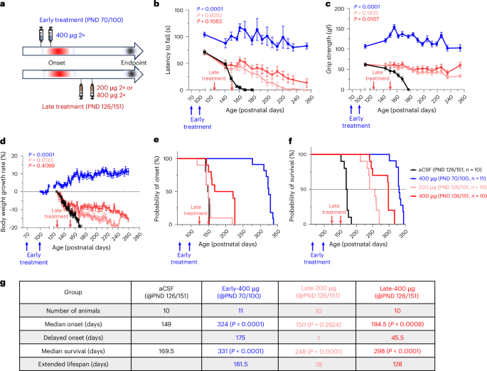
Under freshly prepared 1.2% avertin anesthesia (0.30–0.35 ml/10 g), a 25-gauge needle was stereotaxically placed at 0.2 mm anterior–posterior and 1.0 mm mediolateral (right) from bregma. A total of 10 µl of either aCSF vehicle or RAG-17 was injected into the lateral ventricle at approximately 1 µl s−1. For the early-treatment regimen, SOD1G93A mice received two ICV injections of aCSF or 400 µg RAG-17 on PNDs 70 and 100. For the late-treatment regimen, two ICV injections of 200 or 400 µg RAG-17 were administered on PNDs 126 and 151.
Intrathecal injections in rats
Under 3.0% isoflurane anesthesia, a 30-gauge needle was inserted into the L5–L6 interspace until a tail flick reflex was observed. A 30-µl bolus was injected slowly over 1 min. Female SOD1G93A rats received two intrathecal injections of either NTC or 900 μg RAG-17 on day 0 (PND 70) and day 60 (PND 130). WT littermates received aCSF as a vehicle control. Blinding was applied to the treatment groups.
PK and tissue distribution (liquid chromatography tandem–mass spectrometry)
Rat tissue distribution study
Sprague–Dawley rats (n = 24, 12 animals per sex) received a single intrathecal dose of RAG-17 (1.0 mg/30 μl). Rats were killed at days 1, 7, 28 and 42 (n = 6 per time point, three animals per sex). Brain regions (for example, brain stem, cerebellum, frontal cortex, hippocampus, striatum, cerebrum), spinal cord segments (for example, lumbar, cervical, thoracic) and peripheral tissues (for example, spleen, liver, kidney, lung, heart and muscle) were collected and homogenized on ice (1:9 wt/vol) in lysis buffer (Phenomenex).
Monkey PK study
Forty cynomolgus monkeys (20 animals per sex, ~3 to 4 years of age) were randomized into four groups (five animals per sex per group) receiving either vehicle (aCSF) or RAG-17 at 5, 20 or 50 mg dose−1. Two intrathecal doses were administered on days 1 and 15 via the L4–L5 intervertebral space. CSF (1:1 with stabilizing buffer) was collected at 15 min, day 22 and day 71. Subgroups were killed on day 22 (dosing group; n = 6 per group, equally by sex) and day 71 (recovery group; n = 4 per group, equally by sex). Brain (for example, cerebral cortex) and spinal cord (for example, cervical, thoracic, lumbar) tissues were collected.
Quantification
RAG-17 concentrations were determined using validated methods at WuXi AppTec. The specific assay methods were H59-0049-VM for brain tissue and H59-0050-VM for spinal cord tissue. Data were collected by Analyst and processed using Watson LIMS. The linear dynamic range was 50.0–25,000 ng g−1 (rat tissues) and 20.0–8,000 ng ml−1 (monkey CSF and plasma).
Gene expression analysis (RT–qPCR)
Total RNA was extracted from RNAlater-preserved frozen tissues or cultured cells using either Total RNA Isolation Reagent (Biosharp) or RNAzol (Vigorous). RNA samples were then purified using the RNeasy RNA kit (Qiagen). RT was performed using the PrimeScript RT kit (Takara). The resulting cDNA was amplified in triplicate on a LightCycler 480 real-time PCR system (Roche) using SYBR Premix Ex Taq II (Takara). Amplification was conducted with target-specific primers alongside species-specific internal controls (TBP for human; GAPDH for cynomolgus monkey; primer sequences are provided in Supplementary Information). Primer specificity was verified through melting curve analysis. Relative gene expression was calculated using the ΔΔCt method, and knockdown percentage (%) was determined as (1 − (relative target gene level)) × 100.
Histology, RNAscope and SOD1 ELISA
Histology and immunohistochemistry
L4–L5 rat spinal cord segments were fixed in cold 4% paraformaldehyde, paraffin-embedded and sectioned (4-µm thickness). After antigen retrieval in citrate buffer (pH 6.0, 90 °C), sections were labeled with an anti-ChAT antibody (AB144P, Merck Millipore; 1:400) overnight at 4 °C, followed by incubation with an HRP-conjugated antigoat IgG secondary antibody.
ISH (RNAscope)
After intrathecal administration of RAG-17 (1.6 or 4.8 mg) or saline in Sprague–Dawley rats, brain and spinal cord tissues were collected and paraffin embedded. The biodistribution of the oligonucleotide was analyzed on the tissue sections using the miRNAscope HD Reagent Kit—RED (ACDbio) with a custom-designed antisense probe (ACDbio-1199511-S1) following the manufacturer’s instructions.
Quantification of SOD1 protein
Monkey CSF SOD1 levels were quantified by sandwich ELISA (AE17754MK, Abebio). Samples were incubated in microtiter plates precoated with a capture antibody, followed by sequential incubations with a biotin-conjugated detection antibody and streptavidin-HRP. Optical density was measured at 450 nm (with a correction at 540 nm).
Study design and participants in clinical trial of RAG-17
We performed an open-label, single-center and dose-escalation clinical trial to investigate the safety and tolerability of RAG-17 in patients with ALS with SOD1 gene mutation (CREATION). This clinical trial was approved by the Ethics Committee of Beijing Tiantan Hospital (ethical approval KY2023-015-02) and obtained record-filing approval from the Human Genetic Resources Administration of China. The study was registered with NCT05903690 on 24 May 2023. Written informed consent was obtained from the participants or their legal representatives before enrollment. Six eligible patients from China were enrolled between May 2023 and July 2024. The study was conducted in accordance with the CONSORT framework for transparent reporting of clinical trials.
Patients with SOD1-associated ALS were enrolled based on the following inclusion criteria: (1) deemed medically suitable to complete the trial; (2) aged between 18 and 75 years, irrespective of sex; (3) a confirmed diagnosis of ALS with a documented known SOD1 mutation associated with reported disease progression; (4) FVC of ≥50% predicted value during the screening period; (5) diagnosis of patients meeting the revised El Escorial criteria for confirmed or probable familial or sporadic ALS, as established by the World Federation of Neurology37; (6) provision of written informed consent by the patient or their legal representative and (7) commitment to use effective contraception, with no plans for reproduction or gamete donation during the study and for 3 months thereafter. The exclusion criteria were as follows: (1) carriers of specific SOD1 mutations (nucleotide positions 44–66 from the start of SOD1 protein translation or p.F21C); (2) patients who had previously received or were currently receiving tofersen treatment; (3) a positive test result for human immunodeficiency virus, current or historical; (4) a positive test result for hepatitis C virus antibody, current or historical; (5) active hepatitis B infection (hepatitis B surface antigen positive and/or hepatitis B core antibody positive); (6) the use of other investigational drugs within 30 days or 5 half-lives before screening; (7) contraindications to lumbar puncture; (8) patients with other known diseases related to MN dysfunction that could confound the diagnosis of ALS; (9) a psychiatric disorder diagnosed per Diagnostic and Statistical Manual of Mental Disorders, Fifth Edition (DSM-5) criteria or exhibiting active suicidal ideation; (10) combined with severe liver dysfunction (ALT value or aspartate aminotransferase (AST) value of ≥2.0× the upper limit of normal value), renal dysfunction (serum creatinine of ≥1.5× the upper limit of normal value or estimated glomerular filtration rate of <40 ml min−1 1.73 m−2) or severe heart dysfunction (New York Heart Association Class III or IV); (11) permanent mechanical ventilation dependence; (12) history of alcohol or substance abuse; (13) pregnancy, lactation or intent to conceive; (14) concurrent enrollment in another clinical trial involving an investigational therapy; (15) recent vaccination (within 28 days); (16) contraindications for magnetic resonance imaging (MRI; that is, claustrophobia) and (17) any other condition that may interfere with follow-up.
Clinical procedures and dosing regimen
RAG-17 was provided as a dry powder by Ractigen Therapeutics. Before administration, it was reconstituted to 5 ml with aCSF. Patients received a single injection between the L2 and L5 lumbar vertebrae based on the investigator’s judgment per visit, on days 1, 15, 29, 60, 120, 180 and 240, for a total of six or seven injections. The first three participants initiated treatment at 60 mg, while the subsequent three began at 90 mg. During the dose-escalation phase (dosing every 2 weeks), the dose was increased by 30 mg per subsequent treatment. During the dose-maintenance phase, a fixed dose was administered every 2 months over an 8-month cycle (Fig. 4a).
Clinical outcomes
The primary endpoints included the following safety evaluation measures: (1) incidence of adverse events (AEs) and SAEs within 240 days after RAG-17 treatment; (2) changes in laboratory assessments, vital signs, ALSFRS-R scores and incidence of abnormalities in heart, lung and abdomen examinations, neurological examinations, as well as ECG within 240 days after the first injection of RAG-17. The ALSFRS-R consists of 12 items across four subdomains of bodily function (bulbar, fine motor, gross motor and breathing). Each item is scored on an ordinal scale from 0 (total loss of function) to 4 (no loss of function), yielding total scores ranging from 0 to 48, with higher scores indicating better function38.
The secondary endpoints included (1) changes in CSF SOD1 protein levels at days 15, 29, 60, 120, 180 and 240 from baseline; (2) changes in plasma NfL levels at days 1, 15, 29, 60, 120, 180 and 240 from baseline; (3) PK of RAG-17 in plasma at days 1, 15, 29, 60, 120, 180 and 240; and (4) time to invasive mechanical ventilation or death.
The exploratory endpoints included changes in (1–7) pulmonary function (including FVC), fatigue severity, muscle strength, gait function, modified Norris scale scores, quality of life and clinical staging, at assessed at days 15, 29, 90, 180 and 240; and (8–9) electromyography and 7 T MRI imaging features, assessed at days 29, 90, 180 and 240. Fatigue severity was assessed using the FSS39. Evaluation of gait function included three-dimensional gait analysis, short physical performance battery, tandem walking and the 6-m walk test. Quality of life was estimated using the ALSAQ-40, a five-level version of the EQ-5D-5L, the HAMA and the HAMD. Clinical staging was evaluated using the London ALS Stage40 and Milano–Torino staging41. The electromyographic features evaluated in this study included the composite muscle action potentials, motor unit number index and motor unit size index. All clinical outcomes were adjudicated by the clinical event adjudication committee, which was composed of independent experts with extensive clinical experience.
Laboratory assessments
Laboratory assessments, including complete blood count, basic metabolic panel and CSF biochemistry, were performed at the central laboratory of Beijing Tiantan Hospital. CSF SOD1 (ELISA; BMS222TEN, Thermo Fisher Scientific) and plasma NfL (Ella, ProteinSimple) were quantified in duplicate and triplicate, respectively, at a certified third-party laboratory (United-Power Pharma Tech). CSF NfL was measured retrospectively at Beijing Tiantan Hospital by chemiluminescence immunoassay (Human Neurofilament Light Chain (NfL) Assay Kit, Vazyme).
Protocol amendments
The study protocol underwent four approved amendments. From v1.1 to v1.2 (February–April 2023), to minimize patient burden from lumbar punctures, CSF SOD1 measurement at day 1 (secondary endpoint) was removed, and PK analysis was limited to plasma. From v1.2 to v1.3 (April–June 2023), the PK blood sampling schedule was optimized with more frequent early time points (1, 2, 4, 6, 12 and 48 h instead of 15 min, 4 h, 48 h and 4 days postdose). From v1.3 to v2.0 (June–August 2023), FVC inclusion threshold was lowered from ≥80% to ≥50% of predicted to facilitate enrollment by broadening the eligible population. The dosing regimen was specified–fourth dose of 120 mg, fifth dose of 150–180 mg (maximum 180 mg). From v2.0 to v2.1 (August–October 2023), the primary safety and tolerability evaluation period was extended from 180 to 240 days (aligned with the final dose), with all exploratory endpoint assessments prolonged accordingly. The fourth dose increased from 120 mg to 150 mg. The drug storage condition was revised from −20 ± 5 °C to 2–8 °C. All amendments were approved by the institutional ethics committee before enrolling relevant patients.
Ethical considerations and participant enrollment in clinical trial of RAG-17
This clinical trial was approved by the Ethics Committee of Beijing Tiantan Hospital (KY2023-015-02). Six eligible patients were enrolled from 22 May 2023 to 27 November 2023 in China. Written informed consent was obtained from all participants or their legally authorized representatives before enrollment in accordance with the Declaration of Helsinki. Participants received a fixed travel allowance and a modest reimbursement for blood and CSF collection procedures; no other compensation was provided for study participation. This report incorporates data from all participants who received RAG-17 during the study. The data collection period spanned from 24 May 2023 to 11 July 2024. The final dataset was locked as of 8 October 2024.
Sex and gender considerations
Sex or gender information was collected based on self-reporting. No sex-based or gender-based subgroup analysis was performed because the study objectives were designed independently of sex or gender.
Statistics and reproducibility
Analysis of preclinical data
All statistical analyses were performed using GraphPad Prism software (v11.0.0; 84). Quantitative data are presented as mean ± s.d. or mean ± s.e.m., as indicated in the respective figure legends. For comparisons between two groups, Welch’s t test was used. In specific instances, evaluating a specified direction of change, a one-tailed test was applied. For comparisons among three or more groups, statistical significance was determined by one-way ANOVA followed by Dunnett’s or Tukey’s multiple comparisons test. For data involving multiple groups and time points, a two-way ANOVA followed by Bonferroni’s multiple comparison test was used. Survival and disease-onset data were analyzed using the Kaplan–Meier method, and statistical significance was determined using the log-rank (Mantel–Cox) test. A P value <0.05 was considered statistically significant (*P < 0.05; **P < 0.01; ***P < 0.001; ****P < 0.0001).
Reproducibility
For in vitro assays (for example, serum stability, RT–PCR and cytokine measurements), experiments were performed with biologically independent samples (n = 2 or 3 per group per donor) and were successfully replicated independently at least twice. For in vivo studies, the reported n values represent biologically independent animals, with group sizes ranging from 3 to 20 depending on the experimental endpoint. All histological evaluations, including ISH and ChAT staining, were performed on multiple sections per animal (for example, averaged across two separate sections per animal in the L4–L5 region), and assessments were conducted in a blinded manner to the experimental conditions. Representative images were selected from independent biological replicates (n = 3–20 per group), and the results showed consistent outcomes across replicates.
Study design and sample size
No statistical method was used to predetermine sample size. Sample sizes (for example, n = 10–20 for rodent efficacy models, n = 4–6 for NHPs and n = 3 for in vitro and PK analyses) were chosen based on standard practices in the field to ensure adequate statistical power for evaluating the therapeutic efficacy and PD of RAG-17.
Data exclusion
For the exposure–response correlation analyses in cynomolgus monkeys, samples with tissue concentrations below the limit of quantification were excluded (resulting in n = 23 for spinal cord and n = 11 for brain analyses). Otherwise, no data were excluded from the analyses.
Randomization and blinding
Animals were randomly assigned to treatment groups. Behavioral assessments were performed by investigators blinded to the treatment allocations.
Analysis of clinical data
Given the small sample size and exploratory nature of this study, the analysis was descriptive, and no formal inferential hypothesis testing was performed. Data are presented for all six enrolled participants. No statistical method was used to predetermine the sample size and the investigators were not blinded to allocation during experiments and outcome assessment. No data were excluded from the analyses, and the study was not randomized.
Categorical or ordinal variables (including AEs and SAEs) were described by frequency and percentage, stratified by trial visit and dose cohort. Continuous variables, including biomarker levels (for example, SOD1 and NfL) and clinical scores (for example, ALSFRS-R), are summarized using descriptive statistics (for example, measured concentrations or mean with s.d.) or presented graphically. Specifically, for SOD1 and NfL protein, two types of figures were generated—individual sample concentrations across visits and mean percentage (±s.e.) change from baseline by cohort. Longitudinal changes in clinical scores, including ALSFRS-R, FVC, FSS, gait function, modified Norris scale scores, ALSAQ-40, EQ-5D-5L, HAMA, HAMD and clinical staging during the follow-up period, were described using line charts. Selected laboratory tests, muscle strength, electromyography parameters, specific gait function metrics and the London ALS stage were presented directly as measured values in Supplementary Tables 1–65 and 67–102.
For PK data, individual and mean (±s.e.) concentration–time profiles were plotted by dose cohort. No adjustments were made, and no covariates were formally tested in models due to the descriptive design. Exploratory post hoc analyses were performed, including changes in body weight, CSF protein and NfL, as well as changes in ALSFRS based on baseline disease severity and duration. Statistical analyses were performed with SAS software (v9.4; SAS Institute).
Reporting summary
Further information on research design is available in the Nature Portfolio Reporting Summary linked to this article.



