
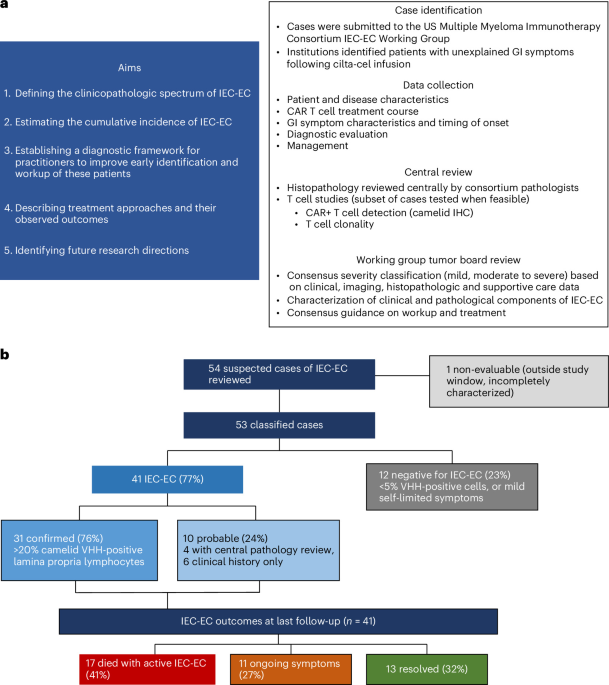
Of 54 suspected cases reviewed, each was discussed at the working group multidisciplinary tumor board and underwent central pathology evaluation when diagnostic tissue was available. Diagnostic confirmation was based on anti-camelid variable heavy-chain (VHH) domain immunohistochemistry (IHC), an assay targeting the camelid-derived binding domain of the cilta-cel CAR construct that identifies CAR+ T cells in tissue sections. Cases were classified as confirmed IEC-EC (≥20% VHH-positive lamina propria lymphocytes), probable IEC-EC (consistent clinical and/or histopathologic features without VHH IHC confirmation) or negative (<5% VHH-positive cells or self-limited symptoms without pathologic evaluation) (Fig. 1b and Table 1; full criteria are found in Methods; severity and outcome definitions are given in Supplementary Tables 1 and 2). No case had a diagnostic biopsy in the indeterminate 5% to 20% VHH IHC range, although levels of camelid VHH between 5% and 20% were seen in biopsies after immunosuppressive therapy initiation and during the treatment course. Anti-camelid VHH IHC was available at five institutions during the study period (Stanford University, MD Anderson Cancer Center, Mayo Clinic, Moffitt Cancer Center and Lombardi Comprehensive Cancer Center). Probable cases most commonly lacked VHH IHC confirmation owing to assay unavailability at the treating institution or lack of diagnostic tissue.
Patient characteristics
The 54 suspected cases of post-cilta-cel GI toxicity were identified across 8 academic institutions between 2019 and 2025. Although the call for cases was inclusive of all BCMA-directed CAR T cell products, only cases following cilta-cel were submitted, potentially reflecting current clinical-use patterns. One patient (patient 25), treated on the CARTITUDE-1 trial (NCT03548207) in 2019, developed diarrhea on day 778, clinically consistent with IEC-EC, with a diagnostic biopsy on day 1,317. The tumor board adjudicated this case non-evaluable and excluded it from the IEC-EC cohort and all denominators for two reasons: it reflected single-site 2019 clinical-trial treatment outside the 2022 to September 2025 real-world window, and its only anti-camelid VHH-stained biopsy (day 1,317) was negative, obtained without confirmed systematic random duodenal and ileal sampling, and was discordant with autopsy-confirmed colitis (tissue not available for camelid-VHH IHC), leaving the case neither confirmed nor confidently negative. The following characteristics describe the 53 evaluable patients.
Across all 53 patients, the median follow-up from cilta-cel infusion was 397 days (interquartile range (IQR), 210–822 days). The median age was 67 years (IQR, 62, 72), with male patients representing 70% of the population. Demographics by diagnostic category are presented in Table 2. Patients received a median of 4 (IQR, 3, 5) previous lines of therapy. High-risk cytogenetics (defined as presence of del(17p), t(4;14) or t(14;16)) were present in 36% of patients and extramedullary disease was present in 15%.
Bridging therapy preceded infusion in 38 of 41 patients with IEC-EC (93%); among 32 evaluable patients, 6 (19%) achieved a partial response or better (Supplementary Figs. 1–3). CAR T cell course and treatment data are summarized in Table 3.
Of the 53 evaluable cases, 12 (23%) were negative for IEC-EC. Ten had GI symptoms prompting endoscopic evaluation, with anti-camelid VHH IHC showing fewer than 5% positive lymphocytes in the lamina propria in all 10 cases. Alternative etiologies were identified in four of these patients, including infectious gastroenteritis (n = 2), bile acid malabsorption (n = 1) and medication toxicity (n = 1); the remaining six patients had self-limited symptoms that resolved without immunosuppressive therapy. Two additional cases without endoscopic evaluation had mild, self-limited symptoms that resolved without intervention. Notably, one patient (patient 52) with fewer than 5% VHH-positive cells carried a separate diagnosis of a CAR+ peripheral T cell lymphoma (PTCL).
Incidence estimation
Extended Data Table 1 details the cumulative incidence calculations. The raw incidence and competing-risk estimates agreed, suggesting that competing mortality had minimal impact on the estimate. Overall cumulative incidence was estimated as follows: (1) raw estimation: 3.0% (95% CI 2.1–4.1%); (2) 12-month competing risk model: 3.0% (95% CI 2.1–4.1%); and (3) sensitivity 12-month competing risk model: 3.0% (95% CI 2.1–4.2%). These estimates derive from a retrospective, referral-based case series assembled across the participating consortium institutions and should be interpreted as consortium-based rather than a definitive population-level incidence. Cumulative incidence showed an overall increase with year-to-year variation, from approximately 2.1% in 2022 to 3.6% from January to September 2025, using the 12-month cumulative incidence estimates. Among the 39 IEC-EC cases through September 2025, 15 (38.5%) were female, 7 (17.9%) identified as Black and 11 (28.2%) as non-White, compared with 43.2%, 10.2% and 20.1%, respectively, among the overall population of 1,308 cilta-cel-treated patients. The raw cumulative incidence of IEC-EC was numerically higher among Black (5.3%) and non-White (4.2%) patients than in the overall population (3.0%). However, chi-square tests of proportions did not find an overrepresentation of female (two sided; χ2 = 0.18; DoF = 1; P = 0.671), Black (two sided; χ2 = 1.70; DoF = 1; P = 0.193) or non-White (two sided; χ2 = 1.07; DoF = 1; P = 0.300) patients with IEC-EC compared with the overall cilta-cel population. No tested variable (peak absolute lymphocyte count (ALC), ferritin and C-reactive protein (CRP), extramedullary disease, high-risk cytogenetics, high marrow plasma-cell burden) differed significantly between IEC-EC and negative cases (Extended Data Table 2).
IEC-EC cohort
Of the 53 evaluable cases, 41 (77%) were adjudicated as IEC-EC. Within the IEC-EC cohort (n = 41), 31 were confirmed by VHH IHC (76%) and 10 were probable (24%), defined as cases with clinical and/or histopathologic features consistent with IEC-EC but without anti-camelid VHH IHC confirmation. Full definitions are included in Methods. Of the ten probable cases, four had pathology available for central review, showing a CD3+ T cell infiltrate on GI tissue biopsy without VHH-confirmation testing. The remaining six probable cases were classified on clinical history alone in the absence of tissue for central pathology review.
The median time from cilta-cel infusion to GI symptom onset in the IEC-EC cohort (n = 41) was 75 days (range 14–541). Diarrhea was the dominant symptom; of the 40 patients with diarrhea, 34 (85%) had a Common Terminology Criteria for Adverse Events grade ≥3 and 2 had low grades (grade 1 or 2); grades were not recorded in 4 patients. One patient presented with abdominal pain without diarrhea.
Laboratory parameters of the 41 patients with IEC-EC after CAR T cell infusion (day 0 to day 30) were as follows: median baseline ferritin was 221.5 ng ml−1 (IQR 82.8–400.3), rising to a peak of 578 ng ml−1 (IQR 299.5–1,318.8) at a median of day 9; median baseline CRP was 2.2 mg dl−1 (IQR 0.6–3.8), rising to 28.4 mg dl−1 (IQR 5.8–56.1) at a median of day 8; and peak ALC was 4.8 × 10⁹ l−1 (IQR 1.9–11.2) at a median of day 13.
Total parenteral nutrition (TPN) was required in 30 patients (73%). At last follow-up, 13 patients (32%) had achieved IEC-EC resolution, 11 (27%) had ongoing symptoms and 17 (41%) had died in the setting of active IEC-EC. Of these 17 deaths, 10 resulted from infectious complications (predominantly septic shock and gram-negative bacteremia; infection was the primary cause in 8), 5 were directly attributable to IEC-EC complications (profound diarrhea, ileus, perforation) and 2 from other causes (embolic cerebrovascular accident and Parkinsonism). One additional patient (patient 41), whose IEC-EC had resolved, subsequently died of unrelated progressive myeloma and was classified as resolved rather than as an IEC-EC-attributable death; all-cause cohort mortality was 18 of 41 (44%). Individual clinical courses are shown in Fig. 2a and characteristics by outcome in Extended Data Table 3. Overall survival for the cohort was 66.7% (95% CI 48.6–79.6%) at 12 months and 52.2% (95% CI 31.9–69.1%) at 24 months (Fig. 2b)
a, Swimmer plot depicting the clinical course of all 41 patients with IEC-EC, grouped by diagnostic certainty (confirmed versus probable). Each horizontal bar represents one patient’s course from cilta-cel infusion. The gray bars indicate the pre-colitis period; the mauve bars indicate active IEC-EC; the teal bars indicate resolution. IEC-EC-directed treatment bars are overlaid in the corresponding color (see key). Infection events are marked by shapes: circles, bacterial; squares, viral; and triangles, fungal or parasitic. X markers indicate death, with cause annotated (infection, IEC-EC, other); the arrows indicate alive at last follow-up. Median time from cilta-cel infusion to IEC-EC onset was 75 days (range 14–541). Corticosteroids include systemic agents and oral budesonide. Ustekinumab in patients 6 and 19 was used as IEC-EC-directed therapy. Patient labels outlined in red mark patients who received treatment for another indication: patient 18 received ustekinumab as maintenance for pre-existing Crohn’s disease (not IEC-EC-directed and not shown); patient 19 received empiric budesonide before IEC-EC onset; patient 20 received dexamethasone and anakinra for immune effector cell-associated hemophagocytic lymphohistiocytosis-like syndrome (IEC-HS), diagnosed on day 19 together with human herpesvirus 6 viremia, before IEC-EC onset; and patient 27 received prophylactic dexamethasone before IEC-EC onset, with GI symptoms of approximately 9 days’ duration identified on admission for delayed neurotoxicity. Patients who died (top) and patients alive at last follow-up (bottom) are plotted separately, each grouped by diagnostic certainty, with independent time axes. TMA, thrombotic microangiopathy; MDS, myelodysplastic syndrome; CVA, cerebrovascular accident. b, Kaplan–Meier estimate of overall survival from cilta-cel infusion for all 41 patients with IEC-EC: 66.7% (95% CI 48.6–79.6%) at 12 months and 52.2% (95% CI 31.9–69.1%) at 24 months. Point estimates with 95% confidence intervals are annotated at 12 months and 24 months; numbers at risk are shown below the axis; the curve is truncated when fewer than 5 patients remain at risk.
Delayed neurological toxicity (DNT) co-occurred in 17 of 41 patients with IEC-EC (41%), higher than the approximately 10% reported among all cilta-cel recipients in real-world data11. DNT presentations included parkinsonism (n = 9), cranial nerve palsies (n = 9) and tremors (n = 1), with two patients experiencing both parkinsonism and facial palsy. Among 14 patients for whom temporal data were available, neurologic symptoms preceded GI onset in 11 (79%) by a median of 48 days (IQR 23–90); in the remaining 3, GI symptoms preceded DNT. In 7 of 11 patients in which neurologic symptoms preceded GI onset, DNT was still active at the time of IEC-EC onset, indicating prolonged overlap rather than sequential resolution of the two toxicities. Patients with DNT had higher median peak post-infusion ALC (n = 17; 8.4 (IQR: 4.4–15.1) × 109 l−1) than those without (n = 23; 2.8 (IQR: 1.3–7.1) × 109 l−1) (two-sided Mann–Whitney U test; U = 105; P = 0.013). DNT was not significantly associated with overall survival (two-sided log-rank test; χ2 = 1.17; DoF = 1; P = 0.28) or progression-free survival (two-sided log-rank test; χ2 = 0.42; DoF = 1; P = 0.52) in exploratory analysis; a small sample size precludes firm conclusions (Extended Data Table 4 and Extended Data Fig. 1).
One patient with confirmed IEC-EC (patient 33) had an unusually complex course: parkinsonism and facial palsy developed on day 154 and resolved by day 170, followed by a diagnosis of CAR+ PTCL on day 377 treated with cyclophosphamide, doxorubicin, vincristine and prednisone with brentuximab vedotin and tazemetostat; IEC-EC onset occurred 48 days later on day 425.
Imaging
Imaging was available (Supplementary Table 3) for review in 38 of 41 IEC-EC cases (92.7%); bowel pathology was identified in 26 (68.4%). Colonic involvement was most common (23/38, 60.5%), followed by small bowel involvement (14/38, 36.8%); both were present in 12 cases. Among complications, pneumatosis intestinalis was present in 10 (26.3%), ileus in 6 (15.8%), pneumoperitoneum in 5 (13.2%), bowel obstruction in 2 (5.3%) and pneumomediastinum in 2 (5.3%).
Histopathologic review
Representative histopathologic images for the 28 cases reviewed at Stanford University are provided in a separate Supplementary Pathology Atlas.
Of 53 cases, 45 (85%) underwent central histopathological review (31 confirmed, 4 probable, 10 negative; Extended Data Table 5) with biopsies performed at a median of 112 days post-infusion. In confirmed and probable IEC-EC cases (n = 35), biopsies were performed between days 34 and 682 post-infusion. Upper endoscopy appeared grossly normal in 9 of 22 evaluable cases (41%). Lower endoscopy appeared normal in 14 of 18 evaluable cases (78%). However, duodenal biopsy (whether isolated or in conjunction with an additional biopsy site) results showed infiltration of lymphocytes on microscopic examination, which was diagnostic in the majority of cases (28 of 34, 82%), with the remaining cases identified from the terminal ileum (n = 3), ileum (n = 1), colon (n = 1) and rectum (n = 1).
Histopathologic evaluation of duodenal biopsies consistently showed features similar to previously published cases1,4,5,6,7,8, with villous blunting, increased apoptotic bodies and a dense infiltrate in the lamina propria of small to slightly enlarged lymphocytes (Extended Data Table 5 and Fig. 3a). Intraepithelial lymphocytes were focally present, but not prominent, and associated eosinophils ranged from inconspicuous to prominent (Fig. 3a). IHC assays showed that the large majority of lymphocytes in the abnormal infiltrate were CD3+ and TCRβ+ T cells. The majority of cases showed a CD4+ immunophenotype (19 of 31, 61%), but 8 of 31 (26%) were CD8+ and 4 of 31 (13%) showed a mixed CD4:CD8 infiltrate; CD4:CD8 predominance was estimated by IHC, with greater than 5:1 or less than 1:5 considered skewed. Of 17 tested cases, 14 (82%) expressed at least one cytotoxic marker (TIA1, perforin or granzyme). Two of the three cytotoxic-negative cases were from one institution (patients 15 and 16), and the third (patient 33) was negative for TIA1 and granzyme B, with perforin not performed. Ki-67 was typically low, with a median of 10% (IQR 5–24.5%). No evidence of Epstein–Barr virus was detected by Epstein–Barr virus-encoded small RNA in situ hybridization in seven tested cases, and CD30 was negative in eight tested cases. Histopathologic and immunophenotypic features of confirmed cases were similar to those of probable cases (Extended Data Table 5 and Supplementary Pathology Atlas); findings in three surgical resection specimens are shown in Supplementary Fig. 4.
a, Representative duodenal biopsy from a patient with confirmed IEC-EC (patient 34, day 72 after infusion). H&E staining shows villous blunting, increased apoptotic bodies and a dense lamina propria infiltrate of small to slightly enlarged lymphocytes. IHC for the anti-camelid VHH domain identifies cilta-cel CAR+ cells throughout the infiltrate, which is CD3+ and TCRβ+ with CD4 predominance, scant CD8+ cells and granzyme B expression in a subset of cells; uniform TRBC1 positivity indicates restricted TRBC1 expression. Each specimen was stained once per antibody; images are representative of the 31 confirmed cases reviewed centrally (Extended Data Table 5). Scale bar (H&E), 100 µm; all images are at the same magnification. b, Summary of diagnostic workup and treatment considerations for IEC-EC derived from this multicenter cohort (n = 41 IEC-EC cases). IEC-EC should be suspected in any cilta-cel recipient with new, unexplained GI symptoms; concurrent infection does not exclude IEC-EC. DNT co-occurred in 41% of cases (17 of 41) and frequently preceded GI symptom onset. Upper endoscopy with duodenal biopsy is recommended as the most reliable detection site, with additional biopsies from the terminal ileum and colon when feasible; endoscopically normal appearance does not exclude IEC-EC. Histopathologic evaluation should assess for a dense lymphocytic infiltrate in the lamina propria. Anti-camelid VHH IHC confirms cilta-cel CAR+ T cell presence. A recommended IHC panel includes CD3, CD4, CD8, CD20, CD56, TCRβ, Ki-67 and cytotoxic markers (TIA1, perforin, granzyme B). T cell clonality should be assessed by TRBC1 IHC and/or molecular testing (TCR PCR or NGS). Treatment considerations: CAR+ T cells appear to be the pathologic effector population. Biologic therapy combined with T cell-directed therapy showed the highest resolution rate in this cohort, although conclusions are limited by the retrospective design and confounding factors. Infection surveillance is critical both at diarrhea onset and throughout IEC-EC treatment given the high risk of coinfections. Most patients required nutritional support, including TPN and close monitoring.
Clonality can be tested through molecular testing or inferred with T cell receptor β-chain constant region 1 (TRBC1) IHC: during T cell receptor gene rearrangement, each αβ T cell randomly selects one of two mutually exclusive β-chain constant regions (TRBC1 or TRBC2), and clonal populations show restricted expression of one isoform, analogous to kappa/lambda light-chain restriction in B cell neoplasms12,13. In this cohort, clonality was assessed via TRBC1 IHC and/or molecular testing (polymerase chain reaction (PCR)) or next-generation sequencing (NGS), reported in Extended Data Table 6 in 22 of 31 confirmed patients with IEC-EC (71%) and 1 of 4 probable IEC-EC cases with pathology (25%). Not all patients underwent both assessments; TRBC1 IHC was evaluable in 15 of 31 confirmed cases and molecular clonality testing was performed in 16 of 31, with partial overlap between the two groups (Extended Data Table 6). TRBC1 expression was restricted in 10 of 15 assessable confirmed cases (67%): 4 showed TRBC1 positivity and 6 showed TRBC1 negativity (indicating TRBC2-restricted expression). Molecular T cell clonality testing by PCR or NGS was performed in 16 of 31 confirmed IEC-EC cases; monoclonal TCR gene rearrangements were detected in 13 (81%), with 1 oligoclonal case and 2 negative results. In probable cases, molecular clonality testing was performed in one of the four with pathology, which showed an equivocal result; TRBC1 IHC was not performed in any probable case. Overall, molecular testing detected a dominant T cell clone in 13 of 17 assessed cases (76%): 13 of 16 confirmed cases (81%), while the single probable case tested showed an equivocal result. Oligoclonality was detected by NGS in one confirmed case (patient 22) that showed mixed TRBC1 expression and a mixed CD4 to CD8 ratio as assessed by IHC; this case showed >20% camelid VHH-positive cells (Supplementary Pathology Atlas). In patient 33, who developed IEC-EC after a CAR+ PTCL, T cell receptor sequencing (clonoSEQ) of three post-infusion tissues, the lymph node involved by PTCL (day 391), the bone marrow (day 500) and a colon biopsy obtained during active IEC-EC (day 523) identified the same three dominant rearrangements, one T cell receptor beta (TRB) and two T cell receptors gamma (TRG), in all three sites (Extended Data Table 7). These clones comprised the majority of the lymph node infiltrate (each 36.5% to 59.3% of total nucleated cells and 80.5% to >99.9% of all TRB or TRG sequences, respectively) and were also detectable in the bone marrow (1.4% to 1.8% of nucleated cells; 10.0% to 14.9% of sequences) and colon (1.7% to 1.9% of nucleated cells; 9.2% to 11.5% of sequences), indicating that the T cells infiltrating the gut shared the dominant clonal identity of the CAR+ PTCL. TRBC1 IHC on the colon biopsy was not skewed, as expected for a small clonal population within a polyclonal background and with two of the three rearrangements at the TRG locus.
Patient 19 harbored an SH2B3L390P mutation (variant allele frequency 26%), previously reported in detail as a case of IEC-EC with clonal CAR+ T cell expansion6. This patient’s diarrhea decreased approximately 2 weeks after initiation of cyclosporine on day 196 post-cilta-cel. Serial duodenal biopsies showed progressive recovery of normal villous architecture and decreased lymphocytic infiltrate (Supplementary Pathology Atlas). Biopsies on days 693 and 821 post-cilta-cel showed only rare scattered camelid VHH-positive cells, and on day 821, only one IEC-EC-associated dominant TCR clone (TRB Db1Jb2-1) was detectable by NGS. Peripheral blood TCR minimal residual disease studies on days 207, 372, 555 and 818 showed progressive decrease in the IEC-EC-associated clone from 2,059 sequences per million nucleated cells (day 207) to <20 sequences per million nucleated cells (day 818; Supplementary Pathology Atlas). In a separate cilta-cel cohort, camelid VHH-positive cells in the bone marrow exceeded 20% of cellularity only transiently within the first month after infusion and were below 5% in the later marrow, liver and lymph node samples (Supplementary Fig. 5), indicating that the dense gut infiltrate in IEC-EC exceeds the expected tissue distribution of cilta-cel-derived cells.
Infectious complications
Infectious complications were frequently observed. In 15 of 41 IEC-EC cases (36.6%), an infection either preceded or coincided with diarrhea onset, encompassing viral (12 events in 8 patients), bacterial (7 events in 6 patients), fungal (1 patient) and other (2 patients, Strongyloides and Blastocystis hominis) (Extended Data Fig. 2a). Following IEC-EC diagnosis, infectious complications occurred in 30 of 41 patients (73.2%), with a high burden of both viral and bacterial infections (Extended Data Fig. 2b). A total of 16 patients experienced more than one post-diagnosis infection, reflecting the combined effects of immunosuppressive therapy and mucosal barrier disruption. The most frequently identified pathogens were norovirus (n = 7 patients) and cytomegalovirus (CMV, 8 events across n = 7 patients), followed by Pseudomonas aeruginosa (4 events across n = 3 patients), adenovirus (4 events across n = 3 patients) and Clostridium difficile (n = 4). Fungal infections occurred in three patients. Infectious complications contributed to the majority of deaths (10 of 17), predominantly gram-negative bacteremia and septic shock, underscoring the importance of balancing disease control with immunosuppression with infection risk.
Treatment
Treatment paradigms (Extended Data Table 8) were not standardized and reflect real-world therapy decisions for an evolving entity.
Four patients received no IEC-EC-directed therapy: patient 42 died with IEC-EC symptoms and progressive MM. Patient 44 died from parkinsonism complications while experiencing IEC-EC symptoms. Patient 36 had ongoing, severe disease complicated by cecal volvulus requiring hemicolectomy with ongoing symptoms at last follow-up. Patient 35 had confirmed IEC-EC by VHH IHC staining in the duodenum with concurrent CMV colitis and was treated with ganciclovir followed by valganciclovir, with complete resolution of all abdominal symptoms to date. One additional patient (patient 7), who had pathologically confirmed CAR+ T cell infiltration, was managed with gut-directed therapy (mesalamine) alone and achieved symptom resolution.
The majority of treated patients required multimodal therapy. Corticosteroids were used in the majority of treated patients but were insufficient as sole therapy: all four patients who received corticosteroids alone died. However, two of these four patients (patients 13 and 46) did not initiate corticosteroids until 306 days and 573 days after symptom onset, and in the remaining two patients (patients 15 and 31), therapy was not escalated despite persistent symptoms.
Biologic therapy (infliximab, vedolizumab and/or ustekinumab) was administered in 24 patients (58.5%); among those receiving biologics without T cell-directed agents (n = 13), mortality was 31% with resolution in 3 (23%) and ongoing symptoms in 6 (46%) (Extended Data Table 8). T cell-directed therapy, including cyclophosphamide, cyclosporine and ruxolitinib, was administered in 19 patients (46.3%). Among patients receiving combined biologic and T cell-directed therapy (n = 11), resolution was observed in 6 (55%) with mortality in 3 (27%) (Extended Data Table 8). Resolution was observed in all patients who received T cell-directed therapy before biologic initiation (n = 3); among those in whom biologics were initiated first with T cell-directed therapy added subsequently (n = 8), resolution was observed in 3 (38%) and 3 died (38%) (Extended Data Table 8).
Patients receiving combination therapy with a T cell-directed agent and biologic agent may have had longer survival time in which to receive and respond to treatment, and sicker patients may have been more likely to receive aggressive multimodal therapy, confounding comparisons between combination therapies. The small subgroup sizes and nonrandomized treatment selection preclude firm conclusions regarding the optimal agents, combinations or sequencing. Consistent with these limitations, an exploratory analysis of overall survival across the five treatment approaches (no IEC-EC-directed therapy, corticosteroids alone, corticosteroids with biologics, corticosteroids with T cell-directed therapy and multimodal therapy) showed no significant difference by log-rank testing (P = 0.82); given the small subgroup sizes, this analysis is not shown to avoid overinterpretation of underpowered data.
The role of ‘maintenance’ therapy to prevent IEC-EC recurrence is unknown and requires further investigation. In this cohort, four patients (9, 11, 19 and 41) had resolution of IEC-EC but were receiving ongoing therapy (range 26–588 days) beyond the IEC-EC resolution date.
Patient progression and death
A total of 5 of 41 patients (12.2%) saw progression of their multiple myeloma (patients 5, 19, 41, 42 and 45). Patients 5 and 19 saw resolution of their IEC-EC before progression (119 days and 390 days between IEC-EC resolution and progression, respectively) while patient 45 progressed while still experiencing active IEC-EC.
Two patients who progressed ultimately died. Patient 41 saw resolution of the IEC-EC symptoms 423 days post-infusion and then progressed on day 632, ultimately dying of progressive disease on day 725. Patient 42 did not see resolution of their IEC-EC before ultimately progressing on day 388 post-infusion and then dying on day 582, with cause of death being multifactorial, including refractory myeloma, IEC-EC and chemotherapy complications.
A total of 16 of 41 patients died without experiencing progression of their multiple myeloma; in 4 of the 16 (25%) died was attributed directly to IEC-EC, in 10 (62.5%) to infection arising during active IEC-EC and in 2 (12.5%) due to other causes (n = 1 embolic CVA, n = 1 parkinsonism).