
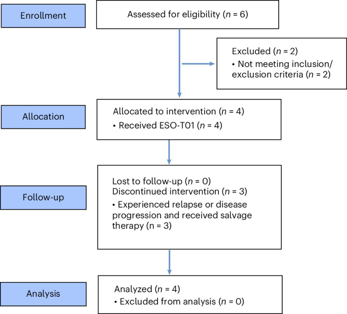
Clinical study design
This phase 1, single-arm, open-label study was conducted at Wuhan Union Hospital, Huazhong University of Science and Technology, China. The aim was to evaluate the safety, tolerability and preliminary efficacy of ESO-T01, a third-generation, non-replicating, self-inactivating lentiviral vector encoding anti-BCMA CAR, in patients with R/R MM. This study was approved by the Ethics Committee of the Union Hospital affiliated to Huazhong University of Science and Technology, Wuhan, China (REC ref no. (2024) 0915-02). The study was registered in ClinicalTrials.gov (NCT06691685) on 9 November 2024. Due to the acquisition of the sponsor, EsoBiotec, by AstraZeneca, the study was terminated, and no more patients were enrolled.
The protocol was amended once to allow prophylactic use of glucocorticoid before infusion, to include patients who suffered from clinical relapse (including new bone lesions or confirmed increase (≥50% increase in sum of the products of the diameters of measurable lesions with an absolute value of ≥1 cm) of lesions or soft‑tissue plasmacytomas), to specify that urine protein electrophoresis, urine immunofixation electrophoresis, and urine free light chain assessments are performed on 24‑h urine collections; and to accept test results obtained within 7 days before screening as valid screening data. The protocol accompanying our previously published report9 was the amended version.
All participants provided written informed consent before any study-related procedures. Participants received study treatment and related procedures free of charge. Reimbursement for transportation and nutrition was provided for each blood collection and follow-up visit.
Sex was assessed via participant self‑report. Due to the small size of the cohort, neither sex nor gender was considered in the enrollment criteria and disaggregated analysis by sex was not performed.
Inclusion criteria
Major inclusion criteria were as follows: age ≥ 18 years, any sex; confirmed diagnosis of MM according to the International Myeloma Working Group criteria with positive BCMA expression on MM cells confirmed by flow cytometry or bone marrow pathology and immunohistochemistry; at least two prior lines of therapy; relapse or disease progression within 12 months before screening; refractoriness to both immunomodulators and proteasome inhibitors; PD during or within 2 months after the most recent antimyeloma therapy; measurable disease as defined as follows; an Eastern Cooperative Oncology Group performance status score of 0–2 and an expected survival of ≥3 months. Measurable disease was defined as serum M protein ≥0.5 g dl−1, or urine M protein ≥200 mg per 24 h, or involved serum free light chain ≥10 mg dl−1 with an abnormal free light chain κ/λ ratio, or clinical relapse (new bone lesions or soft tissue plasmacytomas, excluding osteoporotic fractures, or a confirmed increase of ≥50% in the sum of the products of diameters of measurable lesions, with an absolute value of ≥1 cm, in pre‑existing plasmacytomas or bone lesions).
In addition, patients were required to meet the following criteria at screening or within 2 months before screening: bone marrow function tests showing hemoglobin ≥6 g dl−1 (red blood cell transfusions permitted to maintain this level, and erythropoietin allowed), absolute neutrophil count ≥600 μl−1 (without G‑CSF within 1 week or pegylated G‑CSF within 2 weeks before screening), platelet count ≥50,000 μl−1, lymphocyte count ≥500 μl−1, and absolute CD3+ T cell count ≥150 μl−1; renal function with creatinine clearance ≥45 ml min−1 (Cockcroft–Gault formula); liver function with ALT/AST ≤3.0 × ULN, total bilirubin and ALP ≤2.0 × ULN (except congenital hyperbilirubinemia such as Gilbert’s syndrome, where direct bilirubin ≤1.5 × ULN), and albumin ≥3 g dl−1; cardiac function with left ventricular ejection fraction ≥40%, no clinically important pericardial effusion or electrocardiogram abnormalities; pulmonary function with oxygen saturation ≥90% and no clinically important pleural effusion. Women of childbearing potential must have a negative pregnancy test at screening and before infusion and must not be breastfeeding. Both men and women of childbearing potential must agree to use effective contraception and not to donate germ cells (sperm or oocytes) from the time of signing informed consent until one year after study drug administration. Finally, the subject or a legally authorized representative must provide written informed consent.
Exclusion criteria
Exclusions related to prior treatment comprised: targeted, epigenetic or investigational therapies within 5 half-lives; systemic or cytotoxic therapy within 1 week; proteasome inhibitors or immunomodulators within 2 weeks; radiotherapy within 4 weeks (except when ≤5% of bone marrow reserve was irradiated); allogeneic hematopoietic stem cell transplantation within 6 months or autologous hematopoietic stem cell transplantation within 3 months; prior treatment with a VSVG-pseudotyped virus; and live-attenuated vaccines within 1 month. Disease-related exclusions included active second malignancies (nonmelanoma skin cancer excepted), uncontrolled serious infections, and positive serology for HBV, HCV, HIV or syphilis. Organ dysfunction exclusions were severe heart failure (NYHA class III or IV), myocardial infarction or revascularization within 6 months, clinically important arrhythmias and severe cardiomyopathy. Other exclusions comprised primary immunodeficiency, stroke or seizure within 6 months, dementia, Parkinson’s disease, surgery within 2 weeks, severe allergies to ESO-T01 or tocilizumab, lack of venous access and investigator discretion.
Study endpoints and assessment
The primary endpoints were the safety and tolerability of ESO-T01. Safety parameters included dose-limiting toxicities, adverse events, laboratory tests, vital signs, physical examinations and electrocardiograms. The secondary endpoints were efficacy, pharmacokinetics and pharmacodynamics. Efficacy parameters included time to response, objective response rate, MRD negativity rate, duration of response, progression-free survival, overall survival, objective response rate included sCR, complete response, VGPR and PR. Pharmacokinetic parameters included maximum concentration, time to maximum concentration and area under the curve from time 0 to day 28, for the expansion of BCMA CAR-T cells in peripheral blood after ESO-T01 infusion. Pharmacodynamic parameters included testing of cytokine levels (for example, IL-6, IL-10, IFNγ and TNF), C-reactive protein, ferritin levels, immunoglobulin levels (IgG, IgA, IgM and IgE) and peripheral lymphocyte subpopulations (T, B, NK cell ratios and absolute counts) in peripheral blood after ESO-T01 infusion. The exploratory endpoint was the immunogenicity of ESO-T01 and in vivo generated CAR-T cells by ESO-T01, as well as the presence of shedding of ESO-T01 in blood, saliva, urine and feces. This report focuses on updated safety and efficacy outcomes. The endpoints including safety and tolerability along with early-phase pharmacokinetics or pharmacodynamics profiles have been reported previously in our initial publication on this cohort, and are therefore summarized briefly rather than presented in full here.
Toxicities occurring within 28 days after infusion were recorded as dose-limiting toxicities and graded according to CTCAE v5.0, while CRS and ICANS were graded per the American Society for Transplantation and Cellular Therapy consensus criteria. Beyond 28 days, laboratory abnormalities and other adverse events collected during follow-up visits continued to be graded using CTCAE v5.0. Efficacy evaluation was based on 2016 IMWG criteria.
Lentiviral vector and CAR construct design and production
The CAR backbone of ESO-T01 (PRG1801, WO2025/003526A1 and CN109134665B) consists of an anti-BCMA nanobody (VHH), a CD8α hinge and transmembrane domain, a 4-1BB co-stimulatory domain and a CD3ζ signaling domain, all under the control of a synthetic T cell-specific promoter. To enhance T cell selectivity and reduce off-target transduction, ESO-T01 incorporates additional modifications, including a mutant VSVG envelope, CD47 overexpression, MHC-I knockout and an anti-TCR nanobody. Further details of the vector design, its underlying mechanisms and the Good Manufacturing Practice manufacturing process have been described previously in our earlier report9 and in the study by An et al.40.
Monitoring of CAR-T cells
Surface expression of CAR-transduced T cells in CSF was determined by flow cytometry at the sensitivity of 10−5. Precision count beads (424902; Biolegend) were used for quantification. CSF (1 ml) was incubated with 10 µl counting beads, centrifuged and washed once with PBS. Cells were then incubated with biotinylated human BCMA protein (BC7-H82F0; Acro) for 40 min, washed with PBS, and stained with CD3 APC (317317; Biolegend) and PE-streptavidin (405204; Biolegend) for 15 min. After a final PBS wash, samples were resuspended for flow cytometry analysis. The qPCR was used to detect the vector copy number in four patients as we previously reported9. To better compare the expansion characteristics, CAR copies of in vivo BCMA CAR-T and ex vivo BCMA-GPRC5D CAR-T were detected using ddPCR. Genomic DNA from patient peripheral blood samples was extracted by TIANGEN Genomic DNA Extraction Kit DP340 and 40 ng DNA was used for one test. We used two sets of primers and probes. The first set targets the region spanning 4-1BB and CD3ζ, which is applicable to both in vivo and ex vivo CAR-T products in our study, allowing a better comparison of their differences in CAR-T kinetics. The forward primer was AGAGGAAGATGGCTGTAG; the reverse primer was CTGCTGAACTTCACTCTC; and the probe sequence was FAM-CACATCCTCCTTCTTCTTCTTCTGG-TAMRA (Tsinke, China). The second set targets the anti-BCMA VHH region of the in vivo CAR sequence, enabling specific detection of the kinetics of in vivo CAR-T cells. The forward primer was GGAATCGCTGACGGGTCAGA; the reverse primer was GGCTTCGCTGGGGAACTCA; and the probe sequence was FAM- CTTTGGCTCCTATGGACAGG-TAMRA (General Biol, China). The Bio-Rad CFX96 System was used for quantification.
Immunohistochemistry
Staining for CD38, CD138, CS1 and BCMA was performed on FFPE sections using a Titan automated stainer (Maixin Biotech). Following heat-induced epitope retrieval, sections were processed with a polymer-based detection system and DAB chromogen. Stained slides were digitized and quantified using QuPath v.0.4.3. The catalog of antibodies used for immunohistochemistry are provided in Supplementary Table 2.
Laboratory measurements
Levels of serum creatine kinase, hemoglobin, leukocyte, neutrophil, lymphocyte, platelet, AST, GGT, ferritin, K+, Cl⁻, Ca2+ and immunoglobins were measured in routine laboratory. Flow cytometry was used to analyze the proportion CD4+ T cells, CD8+ T cells, NK cells and B cells within lymphocytes, as well as Tn, Teff, TCM and TEM subsets within CD4+ and CD8+ T cell subpopulations.
T cell phenotype detection
T cell phenotypes were analyzed by flow cytometry. Samples from peripheral blood were stained with the standardized panel: biotinylated human BCMA protein, Fixable Viability Stain 780 (BD, USA), CD3-Percp5.5, CD4-FITC, CD8-PE-Cy7, CD45RA-APC, CD62L-BV510, CD28-BV510, and CD57-PE594, PE-streptavidin. Stained samples were detected using BD FACSymphony A1 (BD) and analyzed using FlowJo software (TreeStar). The clone and catalog of these antibodies are provided in Supplementary Table 3.
Luminex assay
Serum samples were collected at indicated time points and stored at −80 °C until use. The serum of Patient 4 was diluted at a ratio of 1:3, while the serum of the other patients was diluted at a ratio of 1:2. Cytokine levels were measured using a BioPlex Pro Human Cytokine Screening 48-Plex Kit according to the manufacturer’s instructions. Samples were processed on a Luminex 200 instrument, with data acquisition performed by Univ-Bio.
Measurement of vesicular stomatitis virus glycoprotein and CAR antibodies
Anti-VSVG antibodies were detected by ELISA kit (AE-327310-1; Alpha Diagnostic International) according to the manufacturer’s instructions. Plasma samples from healthy donors and patients before and after infusion were collected. The absorbance (optical density value) was measured using Tecan Infinite F50 Microplate Reader. All samples were tested in duplicate wells. To detect anti-CAR antibodies, plasma samples were collected from patients receiving in vivo CAR-T (anti-BCMA VHH), as well as from control groups, including healthy donors, patients with MM receiving ex vivo CAR-T (different anti-BCMA scFv) and patients with ITP receiving ex vivo CAR-T (identical anti-BCMA VHH). For patients with MM, samples were obtained at baseline and after infusion; for patients with ITP, all samples were collected after infusion. Serially diluted BCMA-Fc protein was used as standards. CHO cells expressing BCMA CAR (lentivirus provided by Shenzhen Pregene Biopharma Co., Ltd) were incubated with plasma samples at 4 °C for 40 min, followed by PBS washes and stained with PE anti-human IgG Fc antibody. Serum antibodies concentrations were determined using a standard curve generated by plotting ΔMFI against the log molar concentration of the standards.
Integration site analysis
Lentiviral integration sites were mapped using standardized shearing extension primer tag selection ligation-mediated PCR (S-EPTS/LM-PCR) and next-generation sequencing. Genomic DNA was sheared and amplified with biotinylated primers specific to the viral long terminal repeat. Biotinylated fragments were captured on beads, ligated to barcoded linker, and enriched by nested PCR to incorporate sequencing-specific adapters and secondary barcodes. Final libraries were quantified by Qubit 4.0 and TapeStation 4150, and then sequenced on the MiSeq platform (Illumina)14,41. Data were processed using the Genome Integration Site Analysis pipeline (GENE-IS)42 and R studio (LISA package)43.
Statistical analysis
Data were derived from four patients treated with in vivo CAR-T and control groups (healthy donors, patients with MM treated with ex vivo BCMA CAR-T, and patients with ITP treated with ex vivo BCMA CAR-T). All statistical analyses were performed according to the guideline in section 9 of the study protocol. Sample sizes are as follows: Fig. 2a,b: n = 4 patients; Fig. 2c–f: n = 1 patient (Patient 3, female, 67 years); Fig. 3a–d: n = 4 patients; Fig. 4a: n = 4 patients (in vivo CAR-T) and n = 6 healthy donors; Fig. 4b: n = 4 patients receiving in vivo CAR-T (Day 0 and after infusion), n = 5 patients with MM receiving ex vivo BCMA CAR-T with different anti-BCMA scFv (Day 0 and after infusion), n = 4 patients with ITP treated with CAR-T using the identical anti-BCMA VHH (after infusion) and n = 8 healthy donors; Fig. 4c–f: n = 4 patients; Extended Data Fig. 1a: n = 4 patients: Extended Data Fig. 1b: n = 1 patient (Patient 3, female, 67 years): Extended Data Fig. 2: n = 1 patient (Patient 4, male, 59 years); Extended Data Fig. 3a: n = 1 patient (Patient 1, male, 69 years); Extended Data Fig. 3b: n = 1 patient (Patient 2, female, 40 years); Extended Data Fig. 3c–e: n = 1 patient (Patient 3, female, 67 years); Extended Data Fig. 3f: n = 1 patient (Patient 4, male, 59 years); Extended Data Fig. 4a–c: n = 4 patients; Extended Data Fig. 5a,b: n = 4 patients.
Because of the limited sample size (n = 4), we did not perform formal hypothesis testing, power calculation or inferential statistics. The sample size was not statistically predetermined, no data were excluded and the study was not randomized. The investigators were not blinded to allocation during experiments and outcome assessment. Accordingly, all findings are presented descriptively. Data were analyzed using FlowJo v.10.6.1, GraphPad Prism v.10.4.1 and QuPath v.0.4.3. Additionally, to support reproducibility, the complete raw data and fully annotated analysis templates are available from the corresponding author upon reasonable request, under a standard data transfer agreement to protect participant privacy.
Reporting summary
Further information on research design is available in the Nature Portfolio Reporting Summary linked to this article.



